Quantcast
Channel: ナカライテスク
Viewing all 1104 articles
Browse latest View live

両性イオン性界面活性剤 Fos-Choline®

$
0
0

Affymetrix/Anatrace products社
膜タンパク質研究に適した界面活性剤
両性イオン性界面活性剤 Fos-Choline®

Anatrace_co-brand_logo_4C.jpg

Fos-Choline® は、電荷を持つアミンおよびりん酸基と、アルキル鎖を持つ構造により、両性イオン性界面活性剤の性質を示します。このユニークな構造により、水溶液中で膜タンパク質を安定的に保持できます。また、Fos-Choline® は、ミセルを形成することから、細胞膜からの膜タンパク質の抽出にも利用できます。


界面活性剤の基礎と応用や各製品の物性、保証項目など情報が豊富な、Anatrace2011-2012 カタログのダウンロードが可能です(→PDF 2.9M)。


構造

例)Fos-Choline®-14 の構造


Fos-Choline-14.jpg


CMC(H2O): ~0.12 mM*1(0.0046%)
Aggregation Number(H2O)*2 : ~108
dn/dc(H2O)*2 : 0.1416ml/g
Micelle Size*3 : 47kDa


*1 Anatrace measurement.
*2 Measurement obtained in collaboration with Professor Mark Foster (University of Akron) under an experimental services contract
*3 Strop, P. and Brunger, A. T. (2005) Protein Sci. 14, 2207-2211.


価格表

以下にFos-Choline® のラインアップの中から、一部を紹介します。
ここでは最小容量のみ掲載しています。
*4 Anatrace measurement

製品名CMC*4純度メーカー
製品番号
容量価格オンライン
カタログへ
Fos-Choline®-8, Anagrade

~114mM

(3.4%)

≥99%(HPLC) F300 1 g 55,100 e-Nacalai.jpg
Fos-Choline®-8, Sol-Grade®

~114mM

(3.4%)

≥97%(HPLC) F300S 1 g 44,200 e-Nacalai.jpg
Fos-Choline®-9, Anagrade®

~39.5mM

(1.2%)

≥99%(HPLC) F302 1 g 55,100 e-Nacalai.jpg
Fos-Choline®-9, Sol-Grade®

~39.5mM

(1.2%)

≥97%(HPLC) F302S 1 g 44,200 e-Nacalai.jpg
Fos-Choline®-ISO-9, Anagrade®

~32mM

(0.99%)

≥99%(HPLC) FCI09 1 g 55,100 e-Nacalai.jpg
Fos-Choline®-10, Anagrade®

~11mM

(0.35%)

≥99%(HPLC) F304 1 g 49,200 e-Nacalai.jpg
Fos-Choline®-10, Sol-Grade®

~11mM

(0.35%)

≥97%(HPLC) F304S 1 g 39,300 e-Nacalai.jpg
Fos-Choline®-10, Perdeuterated Head - ≥95%(HPLC) F304PDH 100 mg 97,000 e-Nacalai.jpg
Fos-Choline®-10, Semideuterated Head - ≥95%(HPLC) F304SDH 100 mg 97,000 e-Nacalai.jpg
Fos-Choline®-11, Anagrade®

~1.85mM

(0.062%)

≥99%(HPLC) F306 1 g 55,100 e-Nacalai.jpg
Fos-Choline®-11, Sol-Grade®

~1.85mM

(0.062%)

≥97%(HPLC) F306S 1 g 44,200 e-Nacalai.jpg
Fos-Choline®-11, Perdeuterated Head - ≥95%(HPLC) F306PDH 100 mg 97,000 e-Nacalai.jpg
Fos-Choline®-11, Semideuterated Head - ≥95%(HPLC) F306SDH 100 mg 97,000 e-Nacalai.jpg
Fos-Choline®-ISO-11, Anagrade®

~26.6mM

(0.9%)

≥99%(HPLC) FCI11 1 g 51,800 e-Nacalai.jpg
Fos-Choline®-12, Anagrade®

~1.5mM

(0.047%)

≥99%(HPLC) F308 1 g 49,200 e-Nacalai.jpg
Fos-Choline®-12, Sol-Grade®

~1.5mM

(0.047%)

≥97%(HPLC) F308S 1 g 39,300 e-Nacalai.jpg
Fos-Choline®-12, Deuterated

~1.5mM

(0.047%)

≥90%(HPLC) F308D 100 mg 97,000 e-Nacalai.jpg
Fos-Choline®-12, Perdeuterated Head - ≥95%(HPLC) F308PDH 100 mg 97,000 e-Nacalai.jpg
Fos-Choline®-12, Perdeuterated Tail - ≥95%(HPLC) F308PDT 100 mg 97,000 e-Nacalai.jpg
Fos-Choline®-13, Anagrade®

~0.75mM

(0.027%)

≥99%(HPLC) F310 1 g 55,100 e-Nacalai.jpg
Fos-Choline®-13, Sol-Grade®

~0.75mM

(0.027%)

≥97%(HPLC) F310S 1 g 44,200 e-Nacalai.jpg
Fos-Choline®-14, Anagrade®

~0.12mM

(0.0046%)

≥99%(HPLC) F312 1 g 55,100 e-Nacalai.jpg
Fos-Choline®-14, Sol-Grade®

~0.12mM

(0.0046%)

≥97%(HPLC) F312S 1 g 44,200 e-Nacalai.jpg
Fos-Choline®-14, Deuterated

~0.12mM

(0.0051%)

≥90%(HPLC) F312D 100 mg 99,000 e-Nacalai.jpg
Fos-Choline®-14, Perdeuterated Head - ≥95%(HPLC) F312PDH 100 mg 97,000 e-Nacalai.jpg
Fos-Choline®-14, Semideuterated Head - ≥95%(HPLC) F312SDH 100 mg 97,000 e-Nacalai.jpg
Fos-Choline®-15, Anagrade®

~0.07mM

(0.0027%)

≥99%(HPLC) F314 1 g 55,100 e-Nacalai.jpg
Fos-Choline®-15, Sol-Grade®

~0.07mM

(0.0027%)

≥97%(HPLC) F314S 1 g 44,200 e-Nacalai.jpg
Fos-Choline®-16, Anagrade®

~0.013mM

(0.00053%)

≥99%(HPLC) F316 1 g 55,100 e-Nacalai.jpg
Fos-Choline®-16, Sol-Grade®

~0.013mM

(0.00053%)

≥97%(HPLC) F316S 1 g 44,200 e-Nacalai.jpg

※記載の内容は、'13年10月現在の情報に基づいております。


Santa Cruz社 モノクローナル抗体 新製品

$
0
0

Santa Cruz社では、キナーゼとホスファターゼ、シグナル中間体、転写因子など様々な研究対象用のモノクローナル抗体を新発売しました。


新製品一覧

容量:200μg/ml  価格:51,000円

用途:Western Blotting, Immunoprecipitation, Immunofluorescence, ELISA

略号:Av=Avian(Bird), Bov=Bovine(Cow), Ca=Canine, Hs=Horse(Equine), Hu=Human, Ms=Mouse, Por=Porcine(Pig, Swine), Rt=Rat

製品名交差性抗原アイソタイプメーカー製品番号オンライン
カタログへ
構造タンパク質
CRTAP (E-1) Ms, Hu 171-255 (Hu) mouse IgG1 sc-393136 e-Nacalai.jpg
emerin (H-7) Ms, Rt, Hu 195-224 (Ms) mouse IgG2a sc-393247 e-Nacalai.jpg
KIF17 (B-2) Hu 324-359 (Hu) mouse IgG1 sc-393253 e-Nacalai.jpg
Sarcospan (E-2) Ms, Hu 162-199 (Hu) mouse IgG1 sc-393187 e-Nacalai.jpg
合成と分解
ACAD-10 (F-11) Ms, Rt, Hu, Hs, Bov 409-439 (Hu) mouse IgM sc-393248 e-Nacalai.jpg
ALDH1A2 (G-2) Ms, Rt, Hu, Hs, Ca, Bov, Av 28-51 (Hu) mouse IgG1 sc-393204 e-Nacalai.jpg
Aldolase B (C-11) Ms, Rt, Hu 131-167 (Hu) mouse IgG1 sc-393278 e-Nacalai.jpg
Atg16 (E-10) Hu 15-70 (Hu) mouse IgG1 sc-393274 e-Nacalai.jpg
BUP-1 (B-5) Hu 164-384 (Hu) mouse IgG1 sc-393221 e-Nacalai.jpg
COX6b1 (C-3) Hu 31-80 (Hu) mouse IgG1 sc-393233 e-Nacalai.jpg
CYP27A1 (D-12) Hu 335-451 (Hu) mouse IgG1 sc-393222 e-Nacalai.jpg
DMGDH (E-6) Ms, Rt, Hu, Hs, Ca, Bov 449-483 (Hu) mouse IgG2b sc-393178 e-Nacalai.jpg
MAN1B1 (E-10) Hu 471-657 (Hu) mouse IgG1 sc-393145 e-Nacalai.jpg
mHMGCS (B-8) Ms, Hu 419-488 (Hu) mouse IgG2a sc-393256 e-Nacalai.jpg
NDUFA5 (A-3) Hu 1-116 (Hu) mouse IgG2a sc-393273 e-Nacalai.jpg
Osgep (H-3) Ms, Hu 89-335 (Hu) mouse IgG1 sc-393199 e-Nacalai.jpg
Pancreatic Lipase (G-11) Ms, Rt 29-47 (Rt) mouse IgG2a sc-393158 e-Nacalai.jpg
PLC-XD1 (A-1) Ms, Rt, Hu, Hs, Bov 42-59 (Hu) mouse IgG1 sc-393244 e-Nacalai.jpg
Stim2 (G-3) Ms, Rt, Hu, Hs, Bov, Av 52-73 (Hu) mouse IgM sc-393211 e-Nacalai.jpg
SUMO-2/3/4 (C-3) Hu 1-95 (Hu) mouse IgG1 sc-393144 e-Nacalai.jpg
神経生物学
3PGDH (B-3) Hu 260-533 (Hu) mouse IgG2b sc-393283 e-Nacalai.jpg
ABAT (B-5) Ms, Rt, Hu 59-219 (Hu) mouse IgG1 sc-393142 e-Nacalai.jpg
CNTFRα (F-2) Ms, Rt, Hu 331-360 (Rt) mouse IgM sc-393214 e-Nacalai.jpg
Myelin P2 (A-3) Ms, Hu 18-56 (Hu) mouse IgG2b sc-393173 e-Nacalai.jpg
Neurexophilin-3 (B-11) Ms, Rt, Hu, Hs, Ca 175-202 (Hu) mouse IgG2a sc-393156 e-Nacalai.jpg
NPY1-R (E-4) Ms, Hu 181-271 (Hu) mouse IgG2a sc-393192 e-Nacalai.jpg
NTR1 (G-9) Hu 181-310 (Hu) mouse IgG2b sc-393205 e-Nacalai.jpg
PrP (H-8) Hu 1-253 (Hu) mouse IgG2a sc-393165 e-Nacalai.jpg
rabphilin-3A (D-6) Hu, Rt 516-576 (Hu) mouse IgG2a sc-393197 e-Nacalai.jpg
UIP1 (C-2) Ms 1-300 (Ms) mouse IgG1 sc-393259 e-Nacalai.jpg
輸送関連タンパク質
AP-180 (B-10) Ms, Rt, Hu, Hs, Ca, Bov, Por, Av 2-27 (Ms) mouse IgG1 sc-393266 e-Nacalai.jpg
ASCT1 (C-8) Hu 112-171 (Hu) mouse IgG1 sc-393157 e-Nacalai.jpg
copine I/II/III (E-8) Hu 266-346 (Hu) mouse IgG1 sc-393249 e-Nacalai.jpg
Endophilin B1 (A-9) Hu 250-309 (Hu) mouse IgG1 sc-393191 e-Nacalai.jpg
NIPSNAP1/2 (F-4) Ms, Rt, Hu, Hs, Ca, Bov, Por 197-284 (Ms) mouse IgG1 sc-393201 e-Nacalai.jpg
REEP1 (A-8) Ms, Rt, Hu 145-169 (Hu) mouse IgG1 sc-393242 e-Nacalai.jpg
SCAMP1 (G-8) Ms, Hu 23-93 (Hu) mouse IgG1 sc-393236 e-Nacalai.jpg
Sec61α (G-2) Ms, Rt, Hu, Hs, Ca, Bov, Por, Av 392-423 (Hu) mouse IgG1 sc-393182 e-Nacalai.jpg
キナーゼとホスファターゼ
βPIX (H-3) Ms, Rt, Hu, Ca, Bov 618-646 (Hu) mouse IgG1 sc-393184 e-Nacalai.jpg
ACP1α (A-2) Hu 60-79 (Hu) mouse IgG2a sc-393280 e-Nacalai.jpg
BPNT1 (C-12) Ms, Rt, Hu 64-89 (Hu) mouse IgG1 sc-393185 e-Nacalai.jpg
PIPK II (D-3) Ms, Hu, Ca, Por 2-27 (Hu) mouse IgM sc-393246 e-Nacalai.jpg
PIR1 (B-6) Ms, Rt, Hu, Por 39-58 (Hu) mouse IgM sc-393220 e-Nacalai.jpg
PLEKHM3 (H-2) Ms, Rt, Hu, Hs, Ca, Bov, Av 1-21 (Hu) mouse IgG2a sc-393150 e-Nacalai.jpg
PYK2 (E-3) Ms, Hu, Hs, Ca, Por 980-1009 (Hu) mouse IgG1 sc-393181 e-Nacalai.jpg
Rsk-1 (A-10) Ms, Rt, Hu, Mink, Hs, Ca, Bov, Por 707-735 (Hu) mouse IgG1 sc-393147 e-Nacalai.jpg
SIK2 (B-12) Hu 656-726 (Hu) mouse IgG2a sc-393139 e-Nacalai.jpg
腫瘍抑制因子 / アポトーシス
APIP (G-2) Hu 171-242 (Hu) mouse IgG2a sc-393194 e-Nacalai.jpg
DFNA5 (G-9) Hu 221-496 (Hu) mouse IgG2a sc-393162 e-Nacalai.jpg
DLEC1 (D-11) Ms, Rt, Hu 320-349 (Hu) mouse IgG1 sc-393183 e-Nacalai.jpg
GADD 45γ (B-1) Ms, Hu 95-159 (Hu) mouse IgG2b sc-393261 e-Nacalai.jpg
granzyme M (C-12) Hu 89-126 (Hu) mouse IgG2b sc-393155 e-Nacalai.jpg
MAGE-D4/MAGE-D4B (E-7) Hu 23-310 (Hu) mouse IgG2b sc-393203 e-Nacalai.jpg
ML-IAP (E-3) Hu 72-97 (Hu) mouse IgG1 sc-393237 e-Nacalai.jpg
NRAGE (F-9) Ms, Rt, Hu, Ca, Por 28-57 (Hu) mouse IgG2b sc-393291 e-Nacalai.jpg
Pdcd-2 (H-1) Hu 259-344 (Hu) mouse IgG2a sc-393137 e-Nacalai.jpg
PTEN (F-1) Ms, Rt, Hu, Hs, Ca, Bov, Por, Av 3-29 (Hu) mouse IgG1 sc-393186 e-Nacalai.jpg
Rfp2 (E-6) Hu 212-309 (Hu) mouse IgG2a sc-393257 e-Nacalai.jpg
SESN2 (D-4) Ms, Hu 45-106 (Hu) mouse IgG1 sc-393195 e-Nacalai.jpg
シグナル中間体
14-3-3 ε (F-3) Ms, Rt, Hu, Hs, Ca, Bov, Por, Av 136-175 (Hu) mouse IgG1 sc-393177 e-Nacalai.jpg
Arylsulfatase E (F-4) Hu 513-534 (Hu) mouse IgG2b sc-393224 e-Nacalai.jpg
Ataxin-3 (C-5) Hu 176-228 (Hu) mouse IgG1 sc-393193 e-Nacalai.jpg
BAZ1A (D-5) Hu, Ca, Bov, Por 8-33 (Hu) mouse IgG1 sc-393164 e-Nacalai.jpg
Crk I/II (D-6) Ms, Rt, Hu, Hs, Ca, Bov, Por 109-132 (Hu) mouse IgG1 sc-393160 e-Nacalai.jpg
DcpS (A-12) Ms, Rt, Hu, Hs, Por 97-124 (Hu) mouse IgG2b sc-393226 e-Nacalai.jpg
eIF3K (F-4) Hu, Hs, Ca, Bov, Por 1-218 (Hu) mouse IgG2a sc-393234 e-Nacalai.jpg
FBXO11 (E-9) Ms, Rt, Hu, Hs, Ca, Bov, Por, Av 90-109 (Hu) mouse IgG2b sc-393229 e-Nacalai.jpg
Fyb (H-3) Hu 504-614 (Hu) mouse IgG1 sc-393276 e-Nacalai.jpg
GLB1L2 (B-10) Hu 609-634 (Hu) mouse IgG2b sc-393202 e-Nacalai.jpg
IRS-4 (C-4) Hu 1208-1239 (Hu) mouse IgG1 sc-393207 e-Nacalai.jpg
MAPKAP-1 (F-3) Hu, Hs, Bov 151-450 (Hu) mouse IgG1 sc-393166 e-Nacalai.jpg
MD-1 (H-12) Hu 24-51 (Hu) mouse IgG3 sc-393238 e-Nacalai.jpg
MOB1A (G-7) Ms, Rt, Hu, Ca, Bov 1-64 (Hu) mouse IgG1 sc-393212 e-Nacalai.jpg
NUBPL (G-7) Hu 140-163 (Hu) mouse IgG1 sc-393245 e-Nacalai.jpg
Otospiralin (A-12) Ms 1-89 (Ms) mouse IgG1 sc-393239 e-Nacalai.jpg
PDHA2 (B-3) Ms, Rt 287-312 (Ms) mouse IgG2b sc-393219 e-Nacalai.jpg
Peroxin 1 (D-9) Hu 601-798 (Hu) mouse IgG1 sc-393174 e-Nacalai.jpg
Pinch-1 (B-8) Ms, Hu, Hs, Ca, Bov, Por, Av 12-41 (Hu) mouse IgG1 sc-393151 e-Nacalai.jpg
PRMT2 (B-11) Ms, Rt > Hu 350-376 (Ms) mouse IgM sc-393254 e-Nacalai.jpg
Proteassemblin (B-1) Ms, Rt, Hu, Hs, Ca, Bov 38-63 (Hu) mouse IgG2b sc-393267 e-Nacalai.jpg
Sec5 (F-7) Hu 1-300 (Hu) mouse IgG1 sc-393230 e-Nacalai.jpg
SEMA6D (A-8) Hu 369-420 (Hu) mouse IgG2a sc-393258 e-Nacalai.jpg
SPRED1 (E-5) Hu 135-246 (Hu) mouse IgG1 sc-393198 e-Nacalai.jpg
T6BP (H-6) Hu 387-471 (Hu) mouse IgG1 sc-393143 e-Nacalai.jpg
TRIM (E-12) Ms, Hu 11-197 (Ms) mouse IgG1 sc-393175 e-Nacalai.jpg
TTC30A/B (A-10) Hu 330-523 (Hu) mouse IgG2b sc-393206 e-Nacalai.jpg
WDR1 (B-10) Ms, Hu 425-606 (Hu) mouse IgG2a sc-393159 e-Nacalai.jpg
WSB1 (A-7) Hu, Hs, Ca, Bov, Por 302-354 (Hu) mouse IgG2a sc-393200 e-Nacalai.jpg
ZPBP1 (F-12) Ms, Rt, Hu, Hs 309-338 (Hu) mouse IgG2b sc-393152 e-Nacalai.jpg
細胞膜受容体
Flk-1 (D-8) Ms, Rt, Hu, Hs, Ca, Bov, Por 1282-1321 (Hu) mouse IgG1 sc-393163 e-Nacalai.jpg
Flk-1 (F-10) Ms, Rt, Hu, Hs, Ca, Bov, Por 1292-1321 (Hu) mouse IgG2b sc-393179 e-Nacalai.jpg
GPD1 (H-10) Ms, Hu 300-349 (Hu) mouse IgG1 sc-393161 e-Nacalai.jpg
IL-11Rα (H-3) Ms, Rt 403-432 (Ms) mouse IgG2a sc-393227 e-Nacalai.jpg
IL-3/IL-5/GM-CSFRβ (F-12) Ms, Rt, Hu 867-893 (Ms) mouse IgG1 sc-393281 e-Nacalai.jpg
転写因子
ADAR2 (A-5) Hu 281-370 (Hu) mouse IgG1 sc-393272 e-Nacalai.jpg
CstF-50 (A-5) Ms, Hu, Hs, Ca, Bov, Por 1-300 (Hu) mouse IgG1 sc-393260 e-Nacalai.jpg
DaRS (H-3) Hu 170-467 (Hu) mouse IgG1 sc-393275 e-Nacalai.jpg
Dnmt3b (D-1) Hu 27-54 (Hu) mouse IgG3 sc-393279 e-Nacalai.jpg
ENL (D-6) Hu 131-188 (Hu) mouse IgG1 sc-393196 e-Nacalai.jpg
HCF2 (C-6) Hu 555-792 (Hu) mouse IgG1 sc-393250 e-Nacalai.jpg
HCF2 (D-12) Hu 555-792 (Hu) mouse IgG2b sc-393251 e-Nacalai.jpg
HELIC2 (G-9) Ms, Hu, Hs, Ca, Bov, Por, Av 1-300 (Hu) mouse IgG1 sc-393170 e-Nacalai.jpg
HMG-I/HMG-Y (D-12) Hu 1-95 (Hu) mouse IgG2b sc-393213 e-Nacalai.jpg
ITF-2 (C-1) Ms, Rt, Hu 254-285 (Hu) mouse IgG1 sc-393255 e-Nacalai.jpg
Liprin α2 (C-10) Ms, Hu, Hs, Bov 489-590 (Hu) mouse IgG2a sc-393292 e-Nacalai.jpg
LysRS (B-2) Ms, Rt, Hu, Hs 473-522 (Hu) mouse IgG1 sc-393180 e-Nacalai.jpg
Med30 (C-6) Hu 1-178 (Hu) mouse IgG2a sc-393289 e-Nacalai.jpg
Med7 (E-4) Hu 38-233 (Hu) mouse IgG1 sc-393210 e-Nacalai.jpg
mrnp41 (H-3) Ms, Rt, Hu 12-39 (Hu) mouse IgG2b sc-393252 e-Nacalai.jpg
PATZ1 (H-2) Hu, Hs, Ca, Bov 1-300 (Hu) mouse IgG3 sc-393223 e-Nacalai.jpg
RPUSD3 (C-2) Hu, Ca 92-119 (Hu) mouse IgG2a sc-393209 e-Nacalai.jpg
SCML2 (C-12) Hu 561-597 (Hu) mouse IgM sc-393243 e-Nacalai.jpg
SCML2 (H-3) Hu 551-592 (Hu) mouse IgG2a sc-393265 e-Nacalai.jpg
SMC1α (H-6) Ms, Rt, Hu, Hs, Ca, Bov, Por, Av 1202-1233 (Hu) mouse IgG3 sc-393171 e-Nacalai.jpg
SMC5 (B-11) Hu 901-1090 (Hu) mouse IgG2a sc-393282 e-Nacalai.jpg
SNAI 1 (E-10) Hu 21-150 (Hu) mouse IgG1 sc-393172 e-Nacalai.jpg
SRA (E-5) Ms, Rt 1-220 (Ms) mouse IgG1 sc-393240 e-Nacalai.jpg
TAL1 (B-8) Hu, Ca 272-331 (Hu) mouse IgG2a sc-393288 e-Nacalai.jpg
TAL1 (C-4) Hu, Ca 272-331 (Hu) mouse IgG1 sc-393287 e-Nacalai.jpg
TFIIIC102 (C-2) Ms, Hu 126-413 (Hu) mouse IgG1 sc-393235 e-Nacalai.jpg
Timeless (G-4) Ms, Hu 1-300 (Hu) mouse IgG2a sc-393146 e-Nacalai.jpg
成長因子とホルモン
EMAP II (A-4) Ms, Rt, Hu 268-289 (Hu) mouse IgM sc-393228 e-Nacalai.jpg
Lutropin β (A-9) Ms, Rt 61-103 (Ms) mouse IgG2b sc-393284 e-Nacalai.jpg
PAFAH1B2 (A-6) Ms, Rt, Hu, Hs, Ca, Bov, Por 10-31 (Hu) mouse IgG2a sc-393216 e-Nacalai.jpg
PAFAH1B2 (E-9) Ms, Rt, Hu, Hs, Ca, Bov, Por 10-31 (Hu) mouse IgM sc-393217 e-Nacalai.jpg
Plfr (F-8) Ms 124-244 (Ms) mouse IgG1 sc-393277 e-Nacalai.jpg
リンパ球シグナル伝達
CD3-γ (H-7) Ms, Rt, Hu 151-180 (Hu) mouse IgG3 sc-393271 e-Nacalai.jpg
DOM3Z (B-12) Hu 82-381 (Hu) mouse IgG2a sc-393141 e-Nacalai.jpg
DOM3Z (D-9) Ms, Hu 82-381 (Hu) mouse IgG1 sc-393167 e-Nacalai.jpg
GDP / GTP結合タンパク質
ARAP1 (A-3) Ms, Hu 809-928 (Hu) mouse IgG2b sc-393138 e-Nacalai.jpg
ARL9 (G-10) Ms 51-78 (Ms) mouse IgM sc-393264 e-Nacalai.jpg
E-Ras (B-12) Ms 186-227 (Ms) mouse IgG1 sc-393268 e-Nacalai.jpg
GTPBP9 (F-10) Hu, Hs, Ca, Bov, Por 181-393 (Hu) mouse IgG2a sc-393231 e-Nacalai.jpg
Rab 18 (D-5) Ms, Hu, Hs, Bov, Por 93-206 (Hu) mouse IgG1 sc-393168 e-Nacalai.jpg
Rho GAP p190-B (G-11) Ms, Hu 1091-1250 (Hu) mouse IgG2a sc-393241 e-Nacalai.jpg
Tiam1 (G-1) Ms, Hu 1-300 (Hu) mouse IgM sc-393176 e-Nacalai.jpg
ホメオドメインタンパク質
Nkx-3.1 (A-3) Hu 1-50 (Hu) mouse IgG1 sc-393190 e-Nacalai.jpg
PTF1 (A-3) Ms, Rt, Hu, Ca, Por 291-328 (Hu) mouse IgM sc-393148 e-Nacalai.jpg
細胞接着タンパク質
cadherin-16 (E-7) Ms 1-300 (Ms) mouse IgG1 sc-393153 e-Nacalai.jpg
Integrin α3 (A-6) Hu 841-872 (Hu) mouse IgG1 sc-393290 e-Nacalai.jpg
Integrin β2 (H-7) Hu 1-120 (Hu) mouse IgG1 sc-393140 e-Nacalai.jpg
Laminin γ-2 (G-7) Ms, Rt, Hu, Hs, Ca, Bov 1163-1190 (Hu) mouse IgG2a sc-393225 e-Nacalai.jpg
paxillin (C-10) Ms, Rt, Hu 59-89 (Hu) mouse IgG1 sc-393149 e-Nacalai.jpg
チャネルタンパク質
GABAB R2 (E-4) Ms, Rt, Hu, Ca 183-482 (Hu) mouse IgG1 sc-393286 e-Nacalai.jpg
GABAB R2 (H-10) Ms, Rt, Hu, Ca 183-482 (Hu) mouse IgG2a sc-393270 e-Nacalai.jpg
ROM-K (D-3) Ms, Rt, Hu 321-390 (Hu) mouse IgG2a sc-393189 e-Nacalai.jpg
ステロイド受容体
GR (G-5) Ms, Hu 121-420 (Hu) mouse IgG2b sc-393232 e-Nacalai.jpg
Rev-erbα (E-12) Ms, Hu 21-69 (Hu) mouse IgG1 sc-393215 e-Nacalai.jpg
細胞周期タンパク質
ASF1B (C-6) Hu 1-202 (Hu) mouse IgG2b sc-393169 e-Nacalai.jpg
DHFR (C-4) Ms, Rt, Hu 77-106 (Hu) mouse IgG2b sc-393154 e-Nacalai.jpg
MAD2 (C-9) Ms, Rt, Hu, Hs, Bov, Por 178-205 (Hu) mouse IgG1 sc-393188 e-Nacalai.jpg
NVL (A-10) Hu 405-482 (Hu) mouse IgG2a sc-393285 e-Nacalai.jpg
p-Cdk5 (C-7) Ms, Rt, Hu, Hs, Bov, Por 142-171(Hu) mouse IgG3 sc-377558 e-Nacalai.jpg
PCTAIRE-3 (H-4) Ms 11-69 (Ms) mouse IgG1 sc-393262 e-Nacalai.jpg
その他
C9orf114 (H-7) Ms, Rt, Hu, Hs, Ca, Bov, Por 78-99 (Hu) mouse IgG2a sc-393263 e-Nacalai.jpg
Ste7 (B-12) Saccharomyces cerevisiae 2-24 (S) mouse IgG1 sc-393269 e-Nacalai.jpg

※記載の内容は、'13年12月現在の情報に基づいております。

OriGene社 CRISPR/Cas9ゲノム編集システム

$
0
0

CRISPR/Casはゲノム編集の最新技術として注目されています。高い特異性と低い細胞毒性で、特定のゲノムを切断したり、置換することが可能です。


CRISPR/Cas9ゲノム編集システムは、ヒトU6ポリメラーゼIIIプロモーターから発現されるguide RNAとCas9タンパク質の共発現を必要とします。ゲノム上の標的配列の3'末端側に存在するNGG配列(PAM:protospacer-adjacent motif)を目印として、Cas9は標的配列に相補的なguide RNAと複合体を形成して標的配列を認識し、PAM配列より上流の二本鎖DNAを切断します。切断されたゲノムDNAが修復・組換えされることを利用して、ノックアウトやノックインを作製することができます。


CAS9-Genome-Editing.jpg

OriGene社では、guide RNAとCas9タンパク質を共発現するプラスミドベクターを提供しています。ラインアップとして、設計したguide RNA配列をプラスミドベクターに挿入する方法の違いで、クローニング用に突出末端を持つようにカットされたpre-cut pCas-Guide(pCas-Guide Cloning Kit)、制限酵素BamH I/BsmB Iサイトを持つpCas-Guideの2種類から選択いただけます。


詳細情報 >> OriGene社WEBサイト


この製品に関するご意見・ご照会・お問合せはこちら


メーカー情報 


Cas9 based genome editing has become a popular tool for targeted genome manipulation because of its simplicity and high cutting efficiency. This system requires a functional cas9 protein and a guide RNA for effective double-stranded breakage at a desired site. OriGene has developed the pCas-Guide system, a dual-function vector with both guide RNA and Cas9 expression. OriGene also designed a set of donor cassettes for construction of donor vectors. These include Luciferase-Loxp-Puro-Loxp, tGFP-Loxp-Puro-Loxp and tRFP-Loxp-BSD-Loxp.

 OriGene_CRISPR-Cas9.jpg


  pre-cut pCas-Guide(pCas-Guide Cloning Kit)   


The pCas-Guide vector is designed for cloning a guide RNA insert for genome editing purpose. The vector also has a CMV-driven codon-optimized Cas9. When co-transfected with a proper donor DNA, targeted genome editing can be achieved. The vector retains the ampicillin resistance gene for the selection of E. coli transformants. The vector is supplied as a precut vector, ready for insert ligation. This system has been successfully validated in multiple cases of genome editing.


pre-cut pCas-Guide.jpg 


詳細情報 >> OriGene社WEBサイト


  pCas-Guide    


The pCas-Guide vector is designed for cloning a guide RNA insert for genome editing purpose. Target sequence can be cloned into the vector via BamH I and BsmB I sites. The vector also expresses a CMV-driven codon-optimized Cas9. When co-transfected with a proper donor DNA, targeted genome editing can be achieved. The vector retains the ampicillin resistance gene for the selection of E. coli transformants. 


pCas-Guide.jpg


詳細情報 >> OriGene社WEBサイト


この製品に関するご意見・ご照会・お問合せはこちら  

Tocris社 エピジェネティクス関連製品

$
0
0

tocris-logo-web.jpg


この製品に関するご意見・ご照会・お問合せはこちら


Epigenetics can be defined as acquired changes in chromatin structure that arise independently of a change in the underlying DNA nucleotide sequence. Epigenetic modifications - including acetylation, methylation, phosphorylation, and ubiquitination, amongst others - alter the accessibility of DNA to transcription machinery. Below you will find a sample of Tocris products related to epigenetics.


Cat. No. Product Name Activity
14.3.3 Proteins
2145 Difopein  High affinity inhibitor of 14.3.3 proteins; induces apoptosis
2144 R18 Inhibitor of 14.3.3 proteins; induces apoptosis
Bromodomains
4650 I-BET 151 hydrochloride BET bromodomain inhibitor
4499 (+)-JQ 1  Potent and selective BET bromodomain inhibitor
4445 PFI 1  BET bromodomain inhibitor
DNA Methyltransferases
2624 Decitabine DNA methyltransferase inhibitor
4359 Lomeguatrib MGMT inhibitor
3295 RG 108 Non-nucleoside DNA methyltransferase inhibitor
Histone Acetyltransferases
3084 Anacardic acid  Noncompetitive PCAF/p300 HAT inhibitor
4200 C 646  Selective p300/CBP HAT inhibitor
Histone Deacetylases
4593 GSK J1 Potent histone demethylase JMJD3/UTX inhibitor
4594 GSK J4 Histone demethylase JMJD3/UTX inhibitor; cell permeable
4077 MC 1568 Selectively inhibits HDAC class II (IIa)
3747 NCH 51 Histone deacetylase inhibitor
4403 Pyroxamide Histone deacetylase inhibitor
3810 SBHA Histone deacetylase inhibitor
2682 Sodium 4-Phenylbutyrate Histone deacetylase inhibitor
4270 TC-H 106 Class I histone deacetylase inhibitor
3852 Tranylcypromine hydrochloride Irreversible inhibitor of MAO-A, MAO-B and LSD1
1406 Trichostatin A Histone deacetylase inhibitor
3402 Tubacin HDAC 6 inhibitor; inhibits α-tubulin deacetylation
Histone Methyltransferases
3364 BIX 01294 G9a-like protein and G9a histone lysine methyltransferase inhibitor
4504 Chaetocin Selective histone methyltransferase SUV39H1 inhibitor
3861 UNC 0224 Potent G9a histone lysine methyltransferase inhibitor
4343 UNC 0638  Selective G9a and GLP histone lysine methyltransferase inhibitor
4342 UNC 0646  Potent and selective G9a/GLP inhibitor
Hypoxia Inducible Factors
4408 DMOG Prolylhydroxylase inhibitor
4451 IOX 2 Potent, selective HIF1α prolyl hydroxylase-2 (PHD2) inhibitor
JAK
1571 Cucurbitacin I  Selective inhibitor of STAT3/JAK2 signaling
4338 NSC 33994 JAK2 inhibitor
MBT Domains
4666 UNC 1215  Potent and selective L3MBTL3 domain inhibitor
4516 UNC 926 hydrochloride Binds the MBT domain of L3MBTL1 polycomb group protein
Poly(ADP-ribose) Polymerase
3735 BYK 49187 PARP-1 and PARP-2 inhibitor
1401 NU 1025 Potent PARP inhibitor
3255 PJ 34 hydrochloride Potent PARP inhibitor
3748 XAV 939 Tankyrase inhibitor; inhibits Wnt signaling
Protein Serine/Threonine Phosphatases
1136 Okadaic acid  Protein phosphatase 1 and 2A inhibitor
2613 NSC 87877 Potent inhibitor of shp2 and shp1 PTP

この製品に関するご意見・ご照会・お問合せはこちら

Allele Biotechnology社 黄緑色蛍光タンパク質 mNeonGreen

$
0
0

mNeonGreenは、頭索類Branchiostoma lanceolatum由来の四量体蛍光タンパク質から得られた単量体の黄緑色蛍光タンパク質で、単量体の緑色蛍光タンパク質および黄色蛍光タンパク質の中で最高の輝度を有します。従来のイメージングと同様に、超解像分子イメージングにも優れたタグとして利用できます。また、蛍光共鳴エネルギー移動(FRET)におけるシアン色蛍光タンパク質のアクセプターとして有効です。 


詳細情報 >>


この製品に関するご意見・ご照会・お問合せはこちら


メーカー情報 


For a span of 20 years, researchers have by and large looked to Aequorea victoria Green Fluorescent protein (GFP) for their imaging, and tracking needs. Over this time period, the development of GFP has elicited a wide spectrum of colors and optimized versions for a multitude of applications. As imaging moves into the super resolution era, most common fluorescent proteins, including GFP, may not possess certain characteristics beneficial for super resolution applications. With this in mind, Allele now offers a new high performance monomeric yellow-green fluorescent protein, mNeonGreen, derived from a tetrameric fluorescent protein from the cephalochordate Branchiostoma lanceolatum. mNeonGreen is the brightest monomeric green or yellow fluorescent protein to date, and is an excellent fusion tag for traditional imaging as well as stochastic single-molecule super resolution imaging. Lastly it is a stellar fluorescence resonance energy transfer (FRET) acceptor for cyan fluorescent proteins.  For more information on mNeonGreen's properties view our publication, "A bright monomeric green fluorescent protein derived from Branchiostoma lanceolatum" in the journal Nature Methods.


■ mNeonGreen absorbance, excitation, and emission 

em-ex-graph.png

■ mNeonGreen Basic Properties
Excitation Max (nm) 506
Emission Max (nm) 517
Quantum Yield 0.8
Extinction coefficient 116,000
Brightness 92.8
pKa 5.7


■ Super Resolution Imaging Localization Data 
Fluorescent Protein # Molecules Localized # Photons per Molecule PSF Width (nm)
mNeonGreen 33,783 663 ± 259 167.3 ± 34.1
Clover 15,281 408 ± 263 176.4 ± 57.6
mEGFP 6,803 523 ± 261 165.8 ± 37.1


■ Fluorescence imaging of mNeonGreen fusion vectors

Actin

Plasma Membrane

Microtubules

Replication

actin-color.pngのサムネール画像

actin-binding-color.png

microtubules-color.png replicationfoci-color.png


詳細情報 >>


この製品に関するご意見・ご照会・お問合せはこちら  

Tocris社 2013年12月新製品

$
0
0

tocris-logo-web.jpg


この製品に関するご意見・ご照会・お問合せはこちら


New products are an essential component of our range. Introductions are made on an ongoing basis, with hundreds of new small molecules, peptides and antibodies being added every year. Our aim is to find the latest, otherwise unobtainable research tools, and bring them to the market as quickly as we can.


Cat.No. Product Name Activity
4936 Clozapine N-oxide Clozapine (Cat. No. 0444) metabolite
4869 SR 2211 Selective RORγ inverse agonist
4800 4EGI-1 Inhibitor of eIF4E:eIF4G interaction
4838 AZ 12080282 dihydrochloride Inhibitor of Hedgehog (Hh) signaling
4315 NGD 98-2 hydrochloride High affinity CRF1 antagonist
3567 TC-S 7003 Potent Lck inhibitor
5081 J 147 Neurotrophic compound. Reduces Aβ40 and Aβ42 levels
5062 Cyclotraxin B TrkB receptor antagonist
5056 Ki 16425 LPA1 and LPA3 receptor antagonist
5055 Qc 1 Threonine dehydrogenase inhibitor
4982 ML 337 Selective negative allosteric modulator of mGlu3
4880 ML 281 STK33 inhibitor
4874 SR 1078 RORα/γ agonist
4715 JZL 195 Dual FAAH and MAGL inhibitor
4679 CYM 50358 hydrochloride Potent and selective S1P4 antagonist
4407 TC-G 1004 Potent and selective A2A antagonist
4996 PMX 464 Putative inhibitor of the Trx-TrxR system; antiproliferative
4975 CRT 0066101 Potent protein kinase D (PKD) inhibitor
4954 UAMC 00039 dihydrochloride Dipeptidyl peptidase II (DPP-II) inhibitor
4851 Stoppin 2 Competitive p53-MDM2/MDMX interaction inhibitor


この製品に関するご意見・ご照会・お問合せはこちら

PBL InterferonSource社 VeriKineTM Human Interferon Alpha/Beta Receptor2 ELISA

$
0
0

ヒト IFNAR2 を高感度かつ高い特異性で検出可能!

pbl-web_logo.png


この製品に関するご意見・ご照会・お問合せはこちら


VeriKine™ Human Interferon Alpha/Beta Receptor 2 ELISA Kit

 Product Features and Benefits:

• Provides high sensitivity & excellent specificity for detection to < 5 pg/ml
• Compatible with serum, plasma, cell lysate and cell culture supernatant
• Able to measure the membrane-bound form of IFNAR2 in cell lysates
• No significant cross-reactivity with Human IFNAR1 and analogues
• < 10% inter- and intra-assay CVs
• Conserve high value specimens with low sample requirements (1:100 dilutions of Normal Human Serum (NHS) and Plasma)


Standard_Curve.jpg

This universal ligand-binding receptor for human interferons alpha and beta (Type I IFNs) is directly involved in signal transduction. The receptor is a heterodimer of IFNAR1 and IFNAR2. The IFNAR2 subunit exists in three isoforms, two membrane-bound and one soluble. The soluble isoform has been detected in serum and urine and is reported to be elevated in serum from cancer and autoimmune disease patients. The ability to measure sIFNAR2 in serum can improve understanding of the regulation of the immunomodulatory effects of interferons and perhaps how interferon-based therapies contribute to soluble receptor levels.

The Human Interferon Alpha/Beta Receptor 2 ELISA has been developed to detect and quantitate levels of IFNAR2 to < 5 pg/ml. This new assay provides a novel way to measure human IFNAR2 and will allow scientists to deepen their understanding of interferon-based treatments.


IFNAR2a spike recoveries.jpg

 sIFNAR2.jpg


Product Information:

Catalog Number 41385-1
Description VeriKineTM Human Interferon Alpha/Beta Receptor 2 ELISA
Compatibility Serum, Plasma, Cell Culture Supernatant, Cell Lysate
Package Size 1 x 96-well plate
Assay Range 3.1 - 200 pg/ml

この製品に関するご意見・ご照会・お問合せはこちら


Avanti Polar Lipids社 LIPID MAPS MS解析用脂質標準品

$
0
0

Avanti Polar Lipids社は、LIPID MAPS(LIPID Metabolites And Pathways Strategy)で認可された製造メーカーとして、MS解析用の脂質標準品を供給しています。Quantitative standards(定量分析用標準品)とQualitative standards(定性分析用標準品)の2種類のタイプをラインアップしています。


詳細情報 >> Avanti Polar Lipids社WEBサイト


この製品に関するご意見・ご照会・お問合せはこちら


■ Quantitative standards(定量分析用標準品) 

Glycerophospholipids
12:0-13:0 PC 12:0-13:0 PI
17:0-14:1 PC 17:0-14:1 PI
17:0-20:4 PC 17:0-20:4 PI
21:0-22:6 PC 21:0-22:6 PI
12:0-13:0 PE 13:0 LPC
17:0-14:1 PE 17:1 Lyso PC
17:0-20:4 PE 13:0 LPA
21:0-22:6 PE 17:1 LPA
12:0-13:0 PG 14:1(3)-15:1 CA
17:0-14:1 PG 15:0(3)-16:1 CA
17:0-20:4 PG 22:1(3)-14:1 CA
21:0-22:6 PG 24:1(3)-14:1 CA
12:0-13:0 PS Cardiolipin Mix I
17:0-14:1 PS 17:0-20:4 PI(3)P
17:0-20:4 PS 17:0-20:4 PI(4)P
21:0-22:6 PS 17:0-20:4 PI(5)P
12:0-13:0 PA 17:0-20:4 PI(3,4)P2
17:0-14:1 PA 17:0-20:4 PI(3,5)P2
17:0-20:4 PA 17:0-20:4 PI(4,5)P2
21:0-22:6 PA 17:0-20:4 PI(3,4,5)P3
Sphingolipids
Cer/Sph Mixture I C25 Ceramide
Cer/Sph Mixture II C12 Ceramide-1-Phosphate
Sphingosine (d17:1) Lyso Sphingomyelin (d17:1)
Sphinganine (d17:0) Sphingomyelin (d18:1/12:0)
Sphingosine-1-Phosphate (d17:1) Glucosyl (ß) C12 Ceramide
Sphinganine-1-Phosphate (d17:0) Lactosyl (ß) C12 Ceramide
C12 Ceramide  
Sterols
19:0 Cholesterol ester 24(R/S)-hydroxycholesterol (D6)
cholesterol (D7) 25-hydroxycholesterol (D3)
4ß-hydroxycholesterol (D7) 25-hydroxycholesterol (D6)
6α-hydroxy-5α-cholestanol (D7) 27-hydroxycholesterol (D6)
7α-hydroxycholesterol (D7) 5α,6α-epoxycholesterol (d7)
7ß-hydroxycholesterol (D7) 24(R/S),25-epoxycholesterol (D6)
22(R)-hydroxycholesterol (D7) 7-ketocholesterol (D7)
22(S)-hydroxycholesterol (D7) desmosterol (D6)
Saccharolipid
Kdo2-Lipid A  
Prenols
Nor-Dolichol (13~21)  


■ Qualitative standards(定性分析用標準品) 

Glycerolipids
16:0 MG 1,3-19:0 D5 DG
17:0 MG Tripalmitin
18:1 MG Triheptadecanoate
12:0 DG Triolein (18:1)
16:0 DG Trilinoleate
18:0 DG 14:0-16:1-14:0 TG
1,3-18:1(Trans) DG 1-C15-2,3-18:1 TG
1,3-18:2 DG 16:0-16:0-18:0 TG
18:0-16:0 DG 16:0-16:0-18:1 TG
20:0-18:0 DG 14:0-16:1-14:0 D5 TG
1-C15-3-18:1 DG 15:0-18:1-15:0 D5 TG
1,3-14:0 D5 DG 16:0-18:0-16:0 D5 TG
1,3-15:0 D5 DG 16:0-18:1-16:0 TG
1,3-16:0 D5 DG 17:0-17:1-17:0 D5 TG
1,3-16:1 D5 DG 18:0-18:0-18:1 TG
1,3-17:0 D5 DG 18:1-16:0-18:1 TG
1,3-18:0 D5 DG 19:0-12:0-19:0 D5 TG
1,3-18:1 D5 DG 20:0-20:1-20:0 D5 TG
Glycerophospholipids
16:0 PA 18:0 Lyso PA
16:0-18:1 PA 18:1 Lyso PA
16:0 D31-18:1 PA C16 Lyso PA
16:0-22:6 PA 14:0 Lyso PC
18:0-20:4 PA 16:0 Lyso PC
18:0-22:6 PA 17:0 Lyso PC
16:0 D31-18:1 PC 17:1 Lyso PC
24:0 PC 18:0 Lyso PC
24:1 PC 18:1 Lyso PC
C16-02:0 PC C16 Lyso PC
C16-18:1 PC C18 Lyso PC
C17-02:0 PC C18:1 Lyso PC
C18-02:0 PC C16(Plasm) LPC
C18(Plasm)-18:1 PC C18(Plasm) LPC
14:0 PE 13:0 Lyso PE
15:0 PE 14:0 Lyso PE
16:0 PE 16:0 Lyso PE
17:0 PE 17:1 Lyso PE
18:0 PE 18:0 Lyso PE
18:1 PE (Cis) 18:1 Lyso PE
22:6 PE 13:0 Lyso PG
16:0-18:1 PE 14:0 Lyso PG
16:0 D31-18:1 PE 16:0 Lyso PG
16:0-20:4 PE 17:1 Lyso PG
18:0-20:4 PE 18:0 Lyso PG
18:0-22:6 PE 18:1 Lyso PG
C16-18:1 PE 13:0 Lyso PI
C18(Plasm)-18:1 PE 16:0 Lyso PI
14:0 PG 17:1 Lyso PI
18:0 PG 18:0 Lyso PI
16:0-18:1 PG 18:1 Lyso PI
16:0-18:1 D5 PG 13:0 Lyso PS
16:0 D31-18:1 PG 16:0 Lyso PS
16:0-20:4 PG 17:1 Lyso PS
18:0-18:1 PG 18:0 Lyso PS
18:0-20:4 PG 18:1 Lyso PS
18:0-22:6 PG 14:0 BMP (S,R)
16:0 PI 16:0 BMP (R,S)
18:0 PI 18:1 BMP (S,R)
18:1 PI 16:0-17:0 BMP (R,R)
16:0-18:1 PI 14:0 Hemi BMP (S,R)
16:0 D31-18:1 PI 16:0 Hemi BMP (R,R)
18:0-20:4 PI 17:0 Hemi BMP (R,R)
8:0 PI(3,4)P2 18:1 Hemi BMP (S,R)
8:0 PI(3,5)P2 18:1-18:1-16:0 Hemi BMP (R,R)
8:0 PI(4,5)P2 16:0-16:0-C16:0 Hemi BMP (R,R)
8:0 PI(3,4,5)P3 16:0 BDP (R,R)
16:0 PS 18:1 BDP (S,S)
16:0-18:1 PS 17:0-18:1(3) BDP (R,R)
16:0 D31-18:1 PS 18:1 Succinyl PE
16:0-20:4 PS 18:1 PE-N-19:0
18:0 PS 18:1 PS-N-19:0
18:0-18:1 PS 16:0 Monomethyl PE
18:0-20:4 PS 16:0 Dimethyl PE
18:0-22:6 PS 18:1 DGPP
14:0 Lyso PA 16:0 CDP DG
16:0 Lyso PA 18:1 CDP DG
17:0 Lyso PA  
Sphingolipids
Sphinganine (d18:0) N-C16-deoxysphingosine
12:0(2R-OH) Ceramide N-C24:1-deoxysphingosine
12:0(2S-OH) Ceramide N-C12-deoxysphinganine
16:0(2R-OH) Ceramide N-C16-deoxysphinganine
16:0(2S-OH) Ceramide N-C24:1-deoxysphinganine
17:0(2R-OH) Ceramide N-C12-desoxymethylsphingosine
17:0(2S-OH) Ceramide N-C16-desoxymethylsphingosine
18:0(2R-OH) Ceramide N-C24:1-desoxymethylsphingosine
18:0(2S-OH) Ceramide N-C12-desoxymethylsphinganine
18:1(2R-OH) Ceramide N-C16-desoxymethylsphinganine
18:1(2S-OH) Ceramide N-C24:1-desoxymethylsphinganine
20:0(2R-OH) Ceramide C18 Sphinganine (d18:0)
20:0(2S-OH) Ceramide Sphinganine (d20:0)
22:0(2R-OH) Ceramide C16 Sphinganine-1-Phosphate
22:0(2S-OH) Ceramide C24 Sphinganine-1-Phosphate
24:0(2R-OH) Ceramide C16 Ceramide-1-Phosphate
24:0(2S-OH) Ceramide C24 Ceramide-1-Phosphate
24:1(2R-OH) Ceramide Sphingosine PCholine
24:1(2S-OH) Ceramide Lyso SM (d17:1)
Sphingosine (d18:1) C12 Sphingosyl PE (d17:1)
Sphingosine (d20:1) Phytosphingosine (d17:0)
Sphinganine (d17:0) Glucosyl(ß) C12 Cer
1-deoxysphingosine Glucosyl(ß) C16 Cer
1-deoxysphinganine Glucosyl (ß) C24:1 Ceramide
1-desoxymethylsphinganine Galactosyl (ß) C24:1 Ceramide
1-desoxymethylsphinganine (D5) Lactosyl (ß) C16 Ceramide
N-12:0-1-deoxysphinganine Lactosyl (ß) C24 Ceramide
1-deoxysphinganine D3 3-Sulfo-C12 GalCer
1-deoxy-L-threo-sphinganine D03 3,6-Sulfo-C12 GalCer
N-C12-deoxysphingosine  
Metabolic Intermediates
16:0 aldehyde-d9 16:1 aldehyde-d5
Fatty Acyl Coenzyme A
03:0 Coenzyme A 17:0 Coenzyme A
04:0 Coenzyme A 18:0 Coenzyme A
06:0 Coenzyme A 18:0 α-hydroxy Coenzyme A
08:0 Coenzyme A 19:0 Coenzyme A
10:0 Coenzyme A 20:0 Coenzyme A
12:0 Coenzyme A 21:0 Coenzyme A
13:0 Coenzyme A 22:0 Coenzyme A
14:0 Coenzyme A 23:0 Coenzyme A
14:0 ether Coenzyme A 24:0 Coenzyme A
15:0 Coenzyme A 25:0 Coenzyme A
16:0 Coenzyme A 26:0 Coenzyme A
Sterols
Cholesterol (Ovine Wool) 7-ketocholesterol
Cholesterol (Plant) 7-dehydrocholesterol
zymostenol 7-dehydrocholesterol (d7)
7α,25-dihydroxycholesterol (d6 7α-hydroxycholesterol
7a,25-dihydroxycholesterol(d6) 7α-hydroxycholesterol (D7)
5b,6b-epoxycholestanol (D7) 6αOH (D7) Chol ester
7a,24(R/S)-dihydroxycholesterol (d7) 7αOH (D7) Chol ester
7a-hydroxycholestenone 18:1 Chol (D7) ester
7a-hydroxycholestenone (D7) 7ß-hydroxy-cholesterol
7a,24(R/S)-dihydroxycholestenone (d7) 6α-hydroxy-5α-cholestanol
desmosterol 25-hydroxycholesterol
desmosterol (D6) ester 27-hydroxycholesterol
14-demethyl-lanosterol 27-hydroxycholesterol (D6)
14-demethyl-lanosterol (D6) lanosterol (D6)
stigmasterol dihydrolanosterol (D7)
zymosterol campesterol(d6)
zymosterol (D5) sitosterol(d6)
4ß-hydroxycholesterol cholestanol
5α,6α-epoxycholestanol cholestanol (D5)
5ß,6ß-epoxycholestanol cholestenone
Cholesteryl Esters
14:0 cholesteryl ester 18:0 cholesteryl ester
15:0 cholesteryl ester 20:0 cholesteryl ester
16:0 cholesteryl ester 22:0 cholesteryl ester
17:0 cholesteryl ester  
Anandamides
17:1 ethanolamide  
Prenols
Polyprenol mixture (13~21) Dolichol (13~21)
Polyprenal mixture (13~21) Nor-Dolichoic Acid (13~21)
Fatty Acyl Amides
N,N' 16:1 diacyl L-Lysine N-16:0 L-Ser MeEster
S,N-14:0 Diacyl-L-Cysteine N-16:0 L-Serine
Liponucleotides
17:0-18:1 CDP DG 17:0-18:1 dGDP DG
17:0-18:1 dCDP DG 17:0-18:1 UDP DG
17:0-18:1 ADP-DG 17:0-18:1 dTDP DG
17:0-18:1 dADP DG  


詳細情報 >> Avanti Polar Lipids社WEBサイト


この製品に関するご意見・ご照会・お問合せはこちら


Avanti Polar Lipids社 脂質-タンパク質間相互作用解析用ツール Lipid Snoopers®

$
0
0

Lipid Snoopers®は高純度の脂質がニトロセルロースメンブレン上にスポットされています。製品ラインアップは、ホスファチジルイノシトールでまとめられたInositol Snoopers®、酸化リン脂質でまとめられたOxidized Phospholipid Snoopers®の2種類があります。細胞シグナル伝達関連分子の同定、脂質結合抗体の特性分析、新規脂質結合タンパク質の同定などに利用できます。


詳細情報 >> Avanti Polar Lipids社WEBサイト


この製品に関するご意見・ご照会・お問合せはこちら


メーカー情報 


  Inositol Snoopers®   


Avanti Inositol Snoopers consist of 13 different lipid species spotted individually on a solid support. Each spot contains 1 µg of the highest quality pure lipid and is ideal for investigating lipid-protein interactions. 


Inos-Chart.gif 


Inos_Snooper.gif 


Avanti Snoopers have been used as a method to determine/confirm the specificity of Avanti’s WR304 antibody, which recognizes PIP and PIP2.


Inositol Snoopers were blocked with 3% BSA (fatty acid free) in TBS (0.8% NaCl, 20 mM Tris-HCL pH 7.4) containing 1 mM EDTA. The blocked membranes were then probed with WR304 (1.0 μg/mL) in TBS containing 1% BSA and 1 mM EDTA for 1 h at RT. After washing the membranes 3 times with TBS containing 1 mM EDTA the bound antibody was detected with goat antimouse IgM conjugated to horseradish peroxidase (HRP) (Southern Biotech) at a 1:10,000 dilution in TBS containing 1% BSA and 1 mM EDTA for 1 h at RT. After washing, bound HRP was visualized on an X-ray film with an enhanced chemiluminescence substrate (Pierce). 


詳細情報 >> Avanti Polar Lipids社WEBサイト


  Oxidized Phospholipid Snoopers®    


Avanti Ox-Phospholipid Snoopers consist of 12 different lipid species spotted individually on a solid support. Each spot contains 1 µg of the highest quality pure lipid and is ideal for investigating lipid-protein interactions. 


Ox-Chart.gif 


Ox_Snooper.gif 


Avanti Snoopers have been used as a method to determine/confirm the specificity of Avanti’s E06 antibody, which recognizes the phosphocholine headgroup of oxidized phospholipid that is present in oxidized LDL. E06 does not bind to unoxidized phosphocholine or normal LDL.


Ox-Phospholipid Snoopers were blocked with 3% BSA (fatty acid free) in TBS (0.8% NaCl, 20 mM Tris-HCL pH 7.4). The blocked membranes were then probed with E06 (200 ng/mL) in TBS containing 1% BSA for
1 h at RT. After washing the membranes 3 times with TBS containing
1 mM EDTA the bound antibody was detected with goat antimouse IgM
conjugated to horseradish peroxidase (HRP) (Southern Biotech) at a 1:10,000 dilution in TBS containing 1% BSA for 1 h at RT. After washing, bound HRP was visualized on an X-ray film with an enhanced chemiluminescence substrate (Pierce).


詳細情報 >> Avanti Polar Lipids社WEBサイト


この製品に関するご意見・ご照会・お問合せはこちら  

Polysciences社 酵素固定用レジン・酵素吸着用レジン

$
0
0

Polysciences社では、酵素固定化用のレジンを販売しています。レジンにはアクリル樹脂とスチレン樹脂を採用し、酵素を結合・吸着させる官能基にはエポキシ基、アミノ基、オクタデシル基をそれぞれラインアップしています。担体に固定化された酵素は、操作面では酵素反応後の分離が容易であり、経済面では酵素を繰り返し利用できるなどの利点があります。 


詳細情報 >> Polysciences社WEBサイト


この製品に関するご意見・ご照会・お問合せはこちら


メーカー情報 


Immobilized enzymes are powerful tools to optimize processes in both operative and economic terms.

In addition to a more convenient handling of enzyme preparations, the two main targeted benefits of immobilized enzymes are 1) easy separation of the enzyme from the product, and 2) reuse of the enzyme. Immobilized enzymes allow the easy separation of the catalyst after the reaction e.g. filtration thereby reducing the costs of downstream processing. Moreover, the easy separation of the enzyme from the product simplifies enzyme applications and enables a reliable and efficient reaction technology. On the other hand, the reuse of immobilized enzymes provides cost advantages which are often an essential prerequisite for establishing an enzyme-catalyzed process in the first place. Immobilized enzymes produce important pharmaceuticals and food additives in large ton scale.


Enzyme Immobilization Resins_Kits.jpg


  Epoxy Functionalized   


Epoxy-activated resins are almost ideal matrices to perform easy immobilization of enzymes since they allow multipoint covalent binding between the enzyme and resin.

Epoxy-resins.jpg


  Amine Functionalized   


Another procedure for covalent immobilization of enzymes is based on the use of amino resins. Amino resins can be pre-activated by glutaraldehyde and then used in the covalent immobilization of enzymes (see Figure 4). Reaction of the aldehyde groups with amino groups of enzymes to form Schiff bases is fast and gives stable multipoint covalent binding. An even more stable linkage can be achieved by reduction with borohydrides.

Amino-resins.jpg 


  Resins for Enzyme Adsorption                                                             


This method for the immobilization of enzymes is based on the physical adsorption of enzyme protein on the surface of water-insoluble carriers. The method is very gentle and causes little or no conformational change of the enzyme or destruction of its active center. This method is particularly suitable for applications in organic solvents or hydrophobic media such oils. A major advantage of adsorption as a general method of immobilizing enzymes is that usually no reagents are required.


ECR1091M.jpg Adsorption_C18.jpg


この製品に関するご意見・ご照会・お問合せはこちら  

Tocris社 2014年1月新製品

$
0
0

tocris-logo-web.jpg 


この製品に関するご意見・ご照会・お問合せはこちら


New products are an essential component of our range. Introductions are made on an ongoing basis, with hundreds of new small molecules, peptides and antibodies being added every year. Our aim is to find the latest, otherwise unobtainable research tools, and bring them to the market as quickly as we can.


Cat.No. Product Name Activity
4756 COR 170 Selective CB2 receptor inverse agonist
4452 GGsTop Inhibitor of γ-glutamyl transpeptidase (GGT) 


この製品に関するご意見・ご照会・お問合せはこちら

ペンタフルオロフェニルプロピル基結合型HPLCカラム COSMOSIL® 5PFP

Cytoskeleton社 F-actin蛍光染色試薬 Acti-stain™ Fluorescent Phalloidins キャンペーン

$
0
0

【期  間】

2014年1月1日(水)~2014年2月28日(金)


【内  容】

キャンペーン期間中にご注文いただいた対象製品の偶数本数目
(2本目、4本目、6本目...)を希望納入価格の50%Offでご提供


【対象製品】

・Acti-stain™ 488 phalloidin
・Acti-stain™ 535 (Rhodamine phalloidin)
・Acti-stain™ 555 phalloidin
・Acti-stain™ 670 phalloidin

OriGene社 microRNA(miRNA)発現プラスミドベクター

$
0
0

microRNA(miRNA)は21~23塩基からなる非コードRNAで、多くの生物学的プロセスや疾患の発症に関与することがわかってきています。


OriGene社では、各種miRNAを発現するプラスミドベクターを販売しており、2014年1月20日現在でmiRBase Release 20をカバーするヒト:1829種、マウス:1164、ラット:436種のラインアップを保有しています。プラスミドベクターには、トランスフェクションのモニタリングとしてGFP、セレクションマーカーとしてネオマイシン耐性遺伝子を共発現するpCMV-MIRを採用しています。


詳細情報 >> OriGene社WEBサイト


この製品に関するご意見・ご照会・お問合せはこちら


メーカー情報


MicroRNAs (miRNAs) are naturally occurring non-coding RNAs of 21-23 nt and they have been implicated in many biological processes and disease development. MiRNA are processed in multiple steps:

DNA→ Pri-miRNA → Pre-miRNA→ Mature miRNA

OriGene provides clones for over-expression of miRNA of your choice. A region of ~600bp region encompassing each miRNA is cloned downstream of a CMV promoter. Upon transfection, the cellular machinery will process the CMV-driven expression of miRNA precursor into mature miRNA and cellular function can be analyzed.


  • Genome wide miRNA coverage (miRBase release 20.0)
    - 1829 human, 1164 mouse and 436 rat
  • GFP as reporter for transfection monitoring
  • Neomycin selection marker for stable cell establishment
  • 10 μg transfection-ready DNA using PowerPrep® HP Kits
  • 3'-UTR reporter clones available for miRNA target validation
  • Cloning Vector: pCMV-MIR


pCMV-MIR Vector:

pCMV-MIR.jpg 

All inserts are cloned between SgfI and MluI site 


詳細情報 >> OriGene社WEBサイト


この製品に関するご意見・ご照会・お問合せはこちら


関連製品 


miRNA cDNA synthesis

  • Total RNA isolationg kit
  • miRNA cDNA synthesis kit

  • miRNA detection

  • miRNA Detection Assay
  •  

    Global miRNA profiling
  • qPCR primer Panels
  • qPCR master mix

  • Specific miRNA detection and quantification
  • miRNA primer pairs
  • miRNA copy number standards

  • miRNA expression perturbation
  • miRNA expression plasmids
  • Transfection reagent
  • miRNA targets identification
  • 3' UTR reporter clones
  • Expression cDNA clones

  • Cytoskeleton社 F-actin蛍光染色試薬 Acti-stain™ Fluorescent Phalloidins

    $
    0
    0

    Acti-stain™シリーズは、F-actin(アクチン)の検出に用いられるPhalloidin(ファロイジン)に、高輝度かつ退色耐久性に優れた色素をコンジュゲートした製品です。 


    詳細情報 >> Cytoskeleton社WEBサイト


    ★ キャンペーン実施中 ★

     http://www.nacalai.co.jp/products/campaign/cytoskeleton_acti-stain.html


    この製品に関するご意見・ご照会・お問合せはこちら


    メーカー情報 


    The Acti-stain™ line of fluorescent phalloidins has been developed with an emphasis on creating exceptionally bright and stable probes at an economical price. Side-by-side comparisons with leading competing products demonstrate that you will enjoy savings while not sacrificing the quality of the stain when switching to an Acti-stain™ probe. Additionally, these probes are compatible with popular filter sets such as FITC, TRITC and Cy5. The combination of in-house manufacturing, stringent quality control and convenient packaging provides a great value. Give them a try and see for yourself.


    Product Uses Include

    • Stain F-actin in fixed cells
    • Stabilize actin filaments in vitro
    • Visualize actin filaments in vitro


    Actin staining is very useful in determining the structure and function of the cytoskeleton in living and fixed cells. The actin cytoskeleton is a very dynamic and labile structure in the living cell, but it can be fixed with paraformaldehyde prior to probing or staining for actin structures.


    ■ Biochemical characteristics of fluorescent phalloidins

    Acti-stain.jpg 


      Acti-stain™ 488   


    PHDG1_img1_1.jpg PHDG1_scan.jpg
    Swiss 3T3 cell stained with anti-vinculin (red), Dapi (blue nucleus) and F-actin is stained with Acti-stain™ 488 (green F-actin, Cat.# PHDG1). Absorbance and fluorescence scan of Acti-stain™ 488. Labeled phalloidin was diluted into methanol and its absorbance and excitation spectra were scanned between 350-750 and 500-750 nm, respectively. Absorbance peaks at 500 nm and fluorescence at 550 nm.


      Acti-stain™ 555   


    PHDH1_img1.jpg PHDH1_scan.jpg
    Swiss 3T3 cell with activated Rho, nucleus is stained with Dapi (blue) and F-actin is stained with Acti-stain™ 555 (red F-actin, Cat.# PHDH1). Absorbance and fluorescence scan of Acti-stain™ 555. Labeled phalloidin was diluted into methanol and its absorbance and excitation spectra were scanned between 300-750 and 560-750 nm, respectively. Absorbance peaks at 560 nm and fluorescence at 575 nm.


      Acti-stain™ 670                                       


    PHDN1_img1.jpg PHDN1_scan.jpg
    Mitotic Swiss 3T3 cell, F-actin stained with Acti-stain™ 670 (far-red, Cat. # PHDN1), nuclear DNA stained with Dapi (blue). Absorbance and fluorescence scan of Acti-stain™ 670. Labeled phalloidin was diluted into methanol and its absorbance and excitation spectra were scanned between 300-750 and 600-750 nm, respectively. Absorbance peaks at 625 nm and fluorescence at 675 nm.
     

    詳細情報 >> Cytoskeleton社WEBサイト


    この製品に関するご意見・ご照会・お問合せはこちら  


    InvivoGen社 マイコプラズマの検出から予防・除去

    $
    0
    0

    InvivoGen社
    マイコプラズマの検出から予防・除去


    マイコプラズマ汚染の問題点

    • 実験データの信頼性を損なう恐れ
    • 大切な培養細胞のDNA・RNA・タンパク質合成への影響や染色体の変異を誘発
    • 培養液に明確な変化を引き起こさないため、汚染の継続・拡大が進む

    このようにマイコプラズマによるコンタミネーションは哺乳類細胞を培養する上で重大な問題となっています。
    今回、InvivoGen社が販売しているマイコプラズマの検出試薬、感染予防・除去に関連する製品を紹介します。


    ■ Plasmocin™:マイコプラズマの予防と除去

    ■ Normocin™:マイコプラズマ、細菌、真菌感染の予防

    ■ Primocin™:初代培養細胞用のマイコプラズマ、細菌、真菌感染からの予防

    ■ Plasmocure™:マイコプラズマの除去

    ■ PlasmoTest™:マイコプラズマ検出キット

    ■ マイコプラズマ除去試薬に関する FAQ(よくあるご質問)


    Plasmocin™-マイコプラズマ予防・除去試薬

    Plasmocin™は、非常に好評を頂いているマイコプラズマ予防・除去試薬です。Plasmocin™は、マイコプラズマに作用する強力な2種類の殺菌成分を含有しており、一方はタンパク質合成に作用し、他方はDNA複製に作用します。これら二つの特異的かつ独立したターゲットは、マイコプラズマや他の多くの細菌にのみ見られるもので、真核細胞には全く存在しません。


    特長

    • 2週間でマイコプラズマを除去(Treatment)、マイコプラズマ感染の予防可能(Prophylactic)
    • 細胞内在性のマイコプラズマも除去
    • 動物細胞には作用せず、細胞毒性なし
    • 細胞の代謝への影響は認められない
    • 耐性マイコプラズマが出現しにくい
    • ES細胞・ハイブリドーマ・レトロウイルスパッケージング細胞でも処理実績あり


    推奨使用濃度・使用方法

    Plasmocin™ Treatment(マイコプラズマ除去用)

    推奨使用濃度:25μg/ml(本品1mlで1Lの培地が調製できます。)
    使用方法:
    ①細胞分裂の活発な細胞を12.5-37.5μg/mlのPlasmocin™を含む培地に分注します。
    ②3-4日に一度、新しい培地に交換します(処理期間2週間)。
    ③マイコプラズマフリーの状態を保持するためには、5μg/mlの濃度でPlasmocin™を添加し続けてください。
    (注意)もし2週間処理した後でも除去が不十分だった場合は、37.5μg/mlで引き続き処理してください。


    Plasmocin™ Prophylactic(感染予防用)

    推奨使用濃度:2.5~ 5μg/ml(本品1mlで500ml~1Lの培地が調製できます。)
    使用方法:
    Plasmocin™を細胞培養用培地に添加してください。


    作用ターゲット
    • DNA Gyrase
    • リボソームの50Sサブユニット


    価格表
    製品名製品
    濃度
    メーカー
    製品番号
    製品
    番号
    容量価格オンライン
    カタログへ
    Plasmocin™
    (Treatment)
    25 mg/ml ant-mpt 25993-11 50 mg
    (2x1 ml)
    25,000 e-Nacalai.jpg
    Plasmocin™
    (Prophylactic)
    2.5 mg/ml ant-mpp 25994-01 25 mg
    (10x1 ml)
    17,000 e-Nacalai.jpg

    Normocin™-マイコプラズマ、細菌、真菌感染予防試薬

    Normocin™はマイコプラズマ、細菌および真菌に対して活性のある三つの抗生物質を配合した革新的な抗菌剤です。


    特長

    • マイコプラズマ、細菌、真菌に作用
    • 使用条件下で培養細胞の代謝への影響なし
    • 他の抗菌剤と併用可能


    推奨使用濃度

    100μg/ml(本品1mlで500mlの培地が調製できます。)


    作用ターゲット
    • 原核生物:DNA Gyrase,リボソームの50Sサブユニット
    • 真菌:細胞膜を介したイオン交換


    価格表
    製品名製品
    濃度
    メーカー
    製品番号
    製品
    番号
    容量価格オンライン
    カタログへ
    Normocin™ 50 mg/ml ant-nr-1 14858-15 500 mg
    (10x1 ml)
    26,000

    e-Nacalai.jpg

    ant-nr-2 14858-31 1 g
    (20 ml)
    44,000 e-Nacalai.jpg

    Primocin™-初代培養細胞用のマイコプラズマ、細菌、真菌感染予防試薬

    Primocin™は培養時のコンタミネーションから初代培養細胞を守るために特別に設計された新しい抗菌剤です。 特に解剖の際のコンタミネーションや動物内在性のマイコプラズマから守ります。


    特長

    • マイコプラズマ、細菌、真菌に作用
    • 初代培養細胞に毒性なし
    • 他の抗菌剤は不要


    推奨使用濃度

    100µg/ml(本品1mlで500mlの培地が調製できます。)


    作用ターゲット
    • 原核生物:DNA Gyrase,リボソームの30S、50Sサブユニット
    • 真菌:エルゴステロール


    価格表
    製品名製品
    濃度
    メーカー
    製品番号
    製品
    番号
    容量価格オンライン
    カタログへ
    Primocin™ 50 mg/ml ant-pm-1 14860-65 500 mg
    (10x1 ml)
    30,000

    e-Nacalai.jpg

    ant-pm-2 14860-81 1 g
    (20 ml)
    48,000 e-Nacalai.jpg

    Plasmocure™-マイコプラズマ除去試薬

    非常に稀なケースですが、Plasmocin™に耐性のマイコプラズマが報告されています。これらの耐性マイコプラズマを除去するために、InvivoGen社はPlasmocure™を開発しました。本製品はPlasmocin™とは異なるメカニズムでマイコプラズマに作用する2種類の抗生物質を含有しています。


    特長

    • Plasmocin™に耐性のマイコプラズマを除去可能
    • セルラインに対して微弱な毒性示すが十分な回復期待


    推奨使用濃度

    50µg/ml(本品1mlで2Lの培地が調製できます。)


    作用ターゲット
    • Isoleusyl Synthetase
    • リボソームの50Sサブユニット


    価格表
    製品名製品
    濃度
    メーカー
    製品番号
    製品
    番号
    容量価格オンライン
    カタログへ
    Plasmocure™
    100 mg/ml ant-pc 17837-24 100 mg
    (1x1 ml)
    45,000 e-Nacalai.jpg

    PlasmoTest™-マイコプラズマ検出キット

    従来、マイコプラズマによるコンタミネーションを確かめるには特殊な技術を用いて、定期的に試験を行うほかありませんでした。InvivoGen社が開発したPlasmoTest™は初の人工的に改変された細胞を用いるマイコプラズマ検出キットであり、そのため研究室内で日常的な方法として簡単に確立することができます。


    特長

    • 簡単:特別な装置等は不必要で、基本的な細胞培養知識があればOK
    • 迅速:実質的な操作時間は1 時間以内-オーバーナイトインキュベーション後に検出
    • 多用途:細胞培養液に感染する事でマイコプラズマやアコレプラズマ全種を検出可能
    • 高感度:5×10²-5×10⁵cfu/ml のマイコプラズマを検出可能
    • キット構成:ポジコンおよびネガコンを含むアッセイに必要となる全試薬を含む


    使用方法


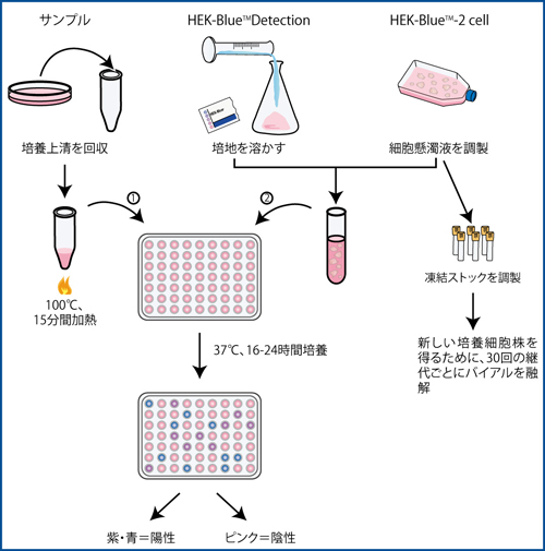
    PlasmoTest_siyouhouhou.jpg


    1. 細胞培養している培地上清を500μlとり、チューブへ移す。
    2. サンプルを100℃、15分間加熱する。
    3. HEK-Blue™ Detection培地を50mlのHEK-Blue™ waterに粉末を溶して調製する。
    4. 96穴プレートにサンプルを50μlずつ入れる。
    5. 96穴プレートにそれぞれのコントロールを50μlずつ入れる。
    6. HEK-Blue™ Detection培地でHEK-Blue™-2cell懸濁液を調製する。
    7. 200μl(~50,000個の細胞)の細胞懸濁液をサンプルまたはコントロールが入っているウェルに添加する。
    8. プレートを37℃のCO2インキュベ ーターで一昼夜(16-24時間)培養する。
    9. 肉眼または620-650nmの分光計でマイコプラズマの有無を検出する。


    キット構成
    • HEK-Blue™-2cells (3-5X10⁶ cells)
    • HEK-Blue™ Selection (2ml) X4
    • Normocin™ (1ml) X4
    • HEK-Blue™ Detection (50ml) X2
    • HEK-Blue™ water (60ml) X2
    • Positive control (死滅しているマイコプラズマ)
    • Negative control

    一度、PlasmoTest™キットをご購入いただくと、後は試薬のみを購入するだけで、さらに試験をすることができます。


    価格表
    製品名メーカー
    製品番号
    製品
    番号
    容量価格オンライン
    カタログへ
    PlasmoTest™
    rep-pt-2 17838-30 1 kit
    (500 samples)
    158,000 e-Nacalai.jpg
    HEK-Blue™
    Selection
    hb-sel 61428-14 5x2 ml 29,000 e-Nacalai.jpg
    Normocin™ ant-nr-1 14858-15 500 mg
    (10x1 ml)
    26,000 e-Nacalai.jpg
    HEK-Blue™
    Detection
    hb-det2 61419-34 5 pouches 47,000 e-Nacalai.jpg
    PlasmoTest™
    Controls
    pt-ctr2 61464-34 200 tests 24,000 e-Nacalai.jpg

    マイコプラズマ除去試薬に関する FAQ(よくあるご質問)

    Q1どのような細胞で処理実績がありますか?
    A1 下記に処理実績のある一部の細胞を掲載します。
    Human Cell Lines:HEK293、HeLa、Colo357、HL60、HSB2、MCF-7、PC3、Hep-G2、MDA-MB231、SW13、SW620、T47D、U-373、U-87
    Rodent Cell Lines:235-1、NIH3T3、B16、CHO、CT60、PC12、C2C12、C6、ES Cells
    その他:Hybridomas、Retrovirus Packaging Cell Line、Bi-color damselfish Cell Lines
    Q2動物細胞に対して毒性はありますか?
    A2 Plasmocin™はマイコプラズマと多くの細菌だけをターゲットとして作用します。細胞に高濃度のPlasmocin™を作用させている場合、細胞の成長率が減速するかもしれませんが、培養液からPlasmocin™を除くと、すぐに正常な成長率に戻ります。
    Q3ペニシリン/ストレプトマイシンを添加した培養液にPlasmocin™、Normocin™、Primocin™を添加できますか?
    A3 Plasmocin™、Normocin™はペニシリン/ストレプトマイシン溶液と互換性があります。ペニシリン/ストレプトマイシンと同時に添加することで、抗菌活性スペクトルを広げることができます。初代培養細胞用のPrimocin™についてはペニシリン/ストレプトマイシンを添加する必要はありません。
    Q4選択用抗生物質存在下でも使用できますか?
    A4 G418、Zeocin™、Hygromycin B、Blasticidin S、Puromycinなどの一般的な選択用抗生物質と互換性があり、同時に使用できます。
    Q5耐性マイコプラズマの出現リスクはありますか?
    A5 耐性菌の存在を確認するために、繰り返し変異の生じる割合を測定しましたが、培養液中に耐性菌は全く出現しませんでした。従って、本製品に対する耐性マイコプラズマは実質的に出現しないと考えられます。もしPlasmocin™耐性マイコプラズマの出現が懸念される場合はPlasmocure™をご使用ください。
    Q6昆虫細胞でも使用できますか?
    A6 哺乳動物細胞と同様のプロトコール(推奨濃度25µg/mlで2週間)でマイコプラズマを除去できます。至適濃度につきましては、50%の範囲で増減(12.5µg/ml~37.5µg/ml)させてお試しください。
    Q7ウイルスパッケージング細胞でも使用できますか?
    A7 同様のプロトコールで使用できます。ウイルス生産前のパッケージング細胞からのマイコプラズマ除去であれば除去用のPlasmocin™ Treatment、ウイルス生産の間であればPlasmocin™ ProphylacticまたはNormocin™をご使用ください。
    Q8tet調節発現系細胞でも使用できますか?
    A8 Plasmocin™はテトラサイクリン・ファミリーに属しませんので、問題なく使用できます。

    ※記載の内容は、'13年11月現在の情報に基づいております。 

    AAT Bioquest社最新ニュースレター:AssayWise Newsletters 2013, Vol 2(2)

    $
    0
    0

    AAT Bioquest社の最新ニュースレターであるAssayWise Newsletters 2013, Vol 2(2) を弊社Web siteへアップしました。

    PDFでご覧いただけます。

    AAT Bioquest社掲載ページはこちら(1ページ目をご覧ください)。


    New Product Highlights

    • Monitoring Cell Cycles
    • Multicolor Labeling of Cell Nucleus with DNA-Binding Dyes
    • DAX-J2™ Ratio 580/460 for Detecting Nitric Oxide (NO)

    Biochemical Assays

    • Neuraminidase Assay
    • Sphingomyelinase Assay
    • Multiplexing Caspase Activity Detection

    Cell-Based Assays

    • Ratio Imaging of Cells with RatioWorks™ Probes
    • Multicolor Labeling of Dead Cells for Flow Cytometric Analysis

    Labeling Probes

    • iFluor™ Fluorescent Probes for Labeling Antibodies
    • trFluor™ Bioconjugates for Developing No Wash ELISA Assays


    AAT-AWL22_200.jpg


    Cytoskeleton社最新ニュースレター:CYTOSKELETON NEWS Jan/Feb 2014

    $
    0
    0

    Cytoskeleton社の最新ニュースレターであるCYTOSKELETON NEWS Jan/Feb 2014を弊社Web siteへアップしました。

    PDFでご覧いただけます。

    CYTOSKELETON NEWS掲載ページはこちら(2ページ目をご覧ください)。


    レビュー
    • Dynein: One Motor, Multiple Neurodegenerative Diseases


     Cytoskeleton_News2014-JanFeb_200.jpg


    「国内研究者使用例」更新

    $
    0
    0

    コンテンツ「国内研究者使用例」に情報を追加しました。


    ■ 今回追加の使用例


    • ナカライテスク Extra PAGE One Precast Gel : MSへの適応
    • ナカライテスク Extra PAGE One Precast Gel : MS/MSへの適応


    • StemCulture社 StemBeads FGF2 : ヒトiPS細胞、ES細胞での使用例


    • ナカライテスク WIDE RANGEゲル調製用緩衝液 : MSへの適応
    • ナカライテスク WIDE RANGEゲル調製用緩衝液 : 大豆タンパク質の分離
    • ナカライテスク WIDE RANGEゲル調製用緩衝液 : マウス脂肪組織タンパク質の分離


    • ナカライテスク Cell Reservoir One(ガラス化法用) : ヒトiPS細胞での使用例


    • Nacalai USA リコンビナント LIF タンパク質 : マウスES細胞での使用例


    • InvivoGen社 Plasmocin™ : Plasmocin™によるマイコプラズマ汚染の検証


    • Biotium社 CF™ Dye CF™ 488A & CF™ 555標識二次抗体の使用例


    国内研究者使用例 一覧へ

    Santa Cruz社 モノクローナル抗体 新製品

    $
    0
    0

    Santa Cruz社では、合成と分解、腫瘍抑制因子 / アポトーシス、シグナル中間体、転写因子など様々な研究対象用のモノクローナル抗体を新発売しました。


    新製品一覧

    容量:200μg/ml  価格:51,000円

    用途:Western Blotting, Immunoprecipitation, Immunofluorescence, ELISA

    略号:Av=Avian(Bird), Bov=Bovine(Cow), Ca=Canine, Hs=Horse(Equine), Hu=Human, Ms=Mouse, Por=Porcine(Pig, Swine), Rt=Rat

    製品名交差性抗原アイソタイプメーカー製品番号オンライン
    カタログへ
    構造タンパク質
    citrin (D-7) Ms, Rt, Hu, Ca, Bov, Por 30-51 (Hu) mouse IgA sc-393303 e-Nacalai.jpg
    Dynactin 2 (G-4) Ms, Hu 102-401 (Hu) mouse IgG1 sc-393389 e-Nacalai.jpg
    Eg5 (D-7) Ms, Rt > Hu 1013-1052 (Ms) mouse IgG1 sc-393311 e-Nacalai.jpg
    KIF17 (F-4) Hu 322-369 (Hu) mouse IgG2b sc-393423 e-Nacalai.jpg
    Misato (F-8) Ms 1-76 (Hu) mouse IgG1 sc-393391 e-Nacalai.jpg
    SLMAP (B-9) Hu 606-828 (Hu) mouse IgG1 sc-393336 e-Nacalai.jpg
    SLMAP (D-3) Hu 606-828 (Hu) mouse IgG2a sc-393393 e-Nacalai.jpg
    Troponin I-SS (A-12) Ms, Rt, Hu 38-58 (Hu) mouse IgM sc-393330 e-Nacalai.jpg
    合成と分解
    ALDH4A1 (C-12) Hu 1-300 (Hu) mouse IgG1 sc-393354 e-Nacalai.jpg
    ALDH4A1 (E-4) Hu 1-300 (Hu) mouse IgG2b sc-393339 e-Nacalai.jpg
    BLVRA (F-1) Hu 75-296 (Hu) mouse IgG1 sc-393385 e-Nacalai.jpg
    CPOX (B-9) Ms, Hu 155-454 (Hu) mouse IgG2a sc-393388 e-Nacalai.jpg
    CYP2U1 (D-4) Hu 9-28 (Hu) mouse IgM sc-393368 e-Nacalai.jpg
    E3BP (F-8) Ms, Hu 172-280 (Ms) mouse IgG1 sc-393449 e-Nacalai.jpg
    NDUFB11 (E-2) Ms 1-151 (Ms) mouse IgG2a sc-393394 e-Nacalai.jpg
    NDUFB3 (F-12) Ms, Hu 1-98 (Hu) mouse IgG2a sc-393351 e-Nacalai.jpg
    NUDT22 (B-2) Ms, Rt, Hu 22-39 (Hu) mouse IgG1 sc-393409 e-Nacalai.jpg
    PDE1B (D-11) Ms, Rt, Hu 435-536 (Hu) mouse IgG1 sc-393349 e-Nacalai.jpg
    PREPL (E-9) Hu 512-727 (Hu) mouse IgG1 sc-393321 e-Nacalai.jpg
    PSMD12 (H-8) Ms, Rt, Hu 3-14 (Hu) mouse IgG2a sc-393401 e-Nacalai.jpg
    神経生物学
    FGGY (C-9) Hu 1-300 (Hu) mouse IgG1 sc-393376 e-Nacalai.jpg
    LIS1 (C-7) Ms, Rt, Hu 1-300 (Hu) mouse IgG1 sc-393320 e-Nacalai.jpg
    MPP4 (C-7) Ms, Hu 165-268 (Ms) mouse IgG1 sc-393413 e-Nacalai.jpg
    VGAT (F-2) Ms, Rt, Hu 71-98 (Hu) mouse IgM sc-393373 e-Nacalai.jpg
    輸送関連タンパク質
    ABCB9 (A-8) Ms > Hu 409-708 (Hu) mouse IgG1 sc-393412 e-Nacalai.jpg
    ACSVL4 (H-6) Ms, Rt, Hu 582-609 (Hu) mouse IgG1 sc-393309 e-Nacalai.jpg
    apoE (E-8) Hu 293-318 (Hu) mouse IgG1 sc-393302 e-Nacalai.jpg
    Synaptotagmin I/II (H-9) Ms, Rt, Hu 301-318 (Hu) mouse IgG1 sc-393392 e-Nacalai.jpg
    キナーゼとホスファターゼ
    AK7 (D-5) Ms, Rt, Hu 518-723 (Hu) mouse IgG1 sc-393337 e-Nacalai.jpg
    ARK-2 (A-3) Hu 48-79 (Hu) mouse IgG2b sc-393357 e-Nacalai.jpg
    eEF2K (B-4) Hu 426-725 (Hu) mouse IgG2a sc-393366 e-Nacalai.jpg
    ERK 3 (G-2) Ms, Rt, Hu 304-339 (Rt) mouse IgG2b sc-393371 e-Nacalai.jpg
    ERK 5 (C-11) Ms, Rt, Hu 786-815 (Hu) mouse IgG1 sc-393405 e-Nacalai.jpg
    h-prune (F-5) Hu 197-249 (Hu) mouse IgG1 sc-393318 e-Nacalai.jpg
    p-PKC δ (A-8) Ms, Rt, Hu Tyr 311 (Hu) mouse IgM sc-377560 e-Nacalai.jpg
    PAK4 (B-6) Ms, Hu 101-240 (Hu) mouse IgG1 sc-393367 e-Nacalai.jpg
    PBK (A-3) Hu 19-252 (Hu) mouse IgG2b sc-393313 e-Nacalai.jpg
    PKN (H-4) Ms, Rt, Hu 923-942 (Hu) mouse IgG1 sc-393344 e-Nacalai.jpg
    PP6 (E-2) Ms, Rt, Hu, Hs, Ca, Bov, Por, Av 266-305 (Hu) mouse IgG1 sc-393294 e-Nacalai.jpg
    SHIP-1 (E-8) Ms, Rt, Hu 1161-1188 (Ms) mouse IgG1 sc-393361 e-Nacalai.jpg
    腫瘍抑制因子 / アポトーシス
    API5 (D-1) Ms, Rt, Hu 72-103 (Hu) mouse IgG1 sc-393341 e-Nacalai.jpg
    caspase-5 p20 (H-2) Hu 120-153 (Hu) mouse IgM sc-393346 e-Nacalai.jpg
    Chibby (C-10) Ms, Rt, Hu 16-37 (Hu) mouse IgG2b sc-393295 e-Nacalai.jpg
    Chibby (G-4) Hu 1-126 (Hu) mouse IgG1 sc-393327 e-Nacalai.jpg
    granzyme M (C-10) Hu 82-129 (Hu) mouse IgG1 sc-393375 e-Nacalai.jpg
    Hemogen (C-7) Ms, Rt, Hu 1-300 (Hu) mouse IgG2a sc-393416 e-Nacalai.jpg
    Hemogen (D-4) Hu 1-300 (Hu) mouse IgG1 sc-393386 e-Nacalai.jpg
    p-CPS2 (F-4) Ms, Rt, Hu, Ca, Bov, Av THuRt 456 (Hu) mouse IgM sc-377559 e-Nacalai.jpg
    p-p53 (F-11) Hu Ser 46 (Hu) mouse IgG1 sc-377561 e-Nacalai.jpg
    ZHX2 (D-2) Hu 578-687 (Hu) mouse IgG2a sc-393399 e-Nacalai.jpg
    シグナル中間体
    β-1,4-GalNAc-T2 (G-1) Ms, Rt, Hu 148-193 (Hu) mouse IgG1 sc-393370 e-Nacalai.jpg
    ANKFY1 (B-6) Hu 1-300 (Hu) mouse IgG1 sc-393353 e-Nacalai.jpg
    calsyntenin-1 (A-4) Hu 647-778 (Hu) mouse IgG2b sc-393383 e-Nacalai.jpg
    CARM1 (G-2) Ms, Rt, Hu 578-608 (Ms) mouse IgG1 sc-393381 e-Nacalai.jpg
    CPSF4 (A-11) Ms, Rt, Hu, Hs, Ca, Bov, Por 1-102 (Hu) mouse IgG1 sc-393316 e-Nacalai.jpg
    CRHSP-24 (D-6) Hu 10-45 (Hu) mouse IgG2a sc-393348 e-Nacalai.jpg
    CTRB1/2 (D-5) Hu 76-123 (Hu) mouse IgG1 sc-393414 e-Nacalai.jpg
    Dvl-2 (D-11) Ms, Rt, Hu 32-75 (Hu) mouse IgG1 sc-393374 e-Nacalai.jpg
    ExoC3L2 (H-5) Hu 97-124 (Hu) mouse IgG1 sc-393332 e-Nacalai.jpg
    FANCG (F-8) Hu 323-622 (Hu) mouse IgG1 sc-393382 e-Nacalai.jpg
    GALK1 (A-2) Ms, Rt, Hu 18-43 (Hu) mouse IgG1 sc-393404 e-Nacalai.jpg
    GALK1 (C-1) Ms, Rt, Hu 18-37 (Hu) mouse IgG2b sc-393356 e-Nacalai.jpg
    GP73 (F-12) Hu 330-364 (Hu) mouse IgG2a sc-393372 e-Nacalai.jpg
    GRP 94 (H-10) Ms, Rt, Hu 200-411 (Hu) mouse IgG2b sc-393402 e-Nacalai.jpg
    Headpin (A-5) Ms, Rt 260-279 (Rt) mouse IgG1 sc-393307 e-Nacalai.jpg
    Headpin (H-7) Ms, Rt 255-282 (Rt) mouse IgG2b sc-393306 e-Nacalai.jpg
    HSPA1L (C-6) Ms 572-637 (Ms) mouse IgG1 sc-393297 e-Nacalai.jpg
    IFIT3 (E-10) Hu 286-490 (Hu) mouse IgG2a sc-393396 e-Nacalai.jpg
    LRFN4 (F-3) Ms, Rt, Hu 209-232 (Hu) mouse IgG3 sc-393425 e-Nacalai.jpg
    NDRG4 (B-10) Ms, Rt, Hu 302-339 (Hu) mouse IgG3 sc-393342 e-Nacalai.jpg
    OPA1 (D-9) Ms, Rt, Hu, Hs, Ca, Bov, Por 647-780 (Hu) mouse IgG1 sc-393296 e-Nacalai.jpg
    PC-1 (H-7) Hu 9-36 (Hu) mouse IgG2b sc-393419 e-Nacalai.jpg
    RWDD4A (D-2) Ms, Hu 101-127 (Hu) mouse IgG3 sc-393410 e-Nacalai.jpg
    SH2-B (E-8) Hu 334-422 (Hu) mouse IgG1 sc-393395 e-Nacalai.jpg
    STAMBP (F-1) Hu > Ms 150-186 (Hu) mouse IgG1 sc-393408 e-Nacalai.jpg
    Sucrase-Isomaltase (A-12) Hu 839-961 (Hu) mouse IgG2a sc-393424 e-Nacalai.jpg
    TBC1D10C (G-5) Hu 26-138 (Hu) mouse IgG1 sc-393379 e-Nacalai.jpg
    TULP3 (H-10) Ms, Rt, Hu 1-300 (Hu) mouse IgG1 sc-393411 e-Nacalai.jpg
    VPS4 (A-11) Ms, Rt, Hu 349-370 (Hu) mouse IgG2b sc-393428 e-Nacalai.jpg
    ZIP1 (F-2) Hu 83-192 (Hu) mouse IgG2a sc-393345 e-Nacalai.jpg
    Zuotin-1 (A-12) Ms, Rt, Hu 10-29 (Hu) mouse IgM sc-393426 e-Nacalai.jpg
    細胞膜受容体
    BMPR-II (E-1) Ms, Rt, Hu, Ca, Bov, Por 61-82 (Hu) mouse IgG1 sc-393304 e-Nacalai.jpg
    IL-2Rα (C-9) Ms, Rt, Hu 23-49 (Ms) mouse IgG2b sc-393326 e-Nacalai.jpg
    IL-2Rβ (C-6) Ms 496-523 (Ms) mouse IgG1 sc-393362 e-Nacalai.jpg
    転写因子
    ADNP (F-5) Ms, Rt, Hu 1-138 (Hu) mouse IgG2a sc-393377 e-Nacalai.jpg
    CD3EAP (A-3) Ms 1-240 (Ms) mouse IgG2a sc-393301 e-Nacalai.jpg
    CHD6 (A-5) Hu 243-320 (Hu) mouse IgG1 sc-393421 e-Nacalai.jpg
    CHRAC17 (D-1) Ms, Hu 1-147 (Hu) mouse IgG1 sc-393397 e-Nacalai.jpg
    COASTER (A-6) Ms, Rt, Hu 983-1010 (Hu) mouse IgG2b sc-393360 e-Nacalai.jpg
    CstF-50 (H-8) Hu, Hs, Ca, Bov, Por 1-300 (Hu) mouse IgG2b sc-393317 e-Nacalai.jpg
    Dicer (E-7) Ms, Rt, Hu 1698-1729 (Hu) mouse IgG2a sc-393328 e-Nacalai.jpg
    DP-1/2 (G-3) Hu 171-237 (Hu) mouse IgG1 sc-393398 e-Nacalai.jpg
    EF-1 γ (C-7) Hu 204-384 (Hu) mouse IgG1 sc-393378 e-Nacalai.jpg
    Eme1 (A-9) Ms 37-91 (Ms) mouse IgG1 sc-393363 e-Nacalai.jpg
    HDAC10 (E-2) Ms, Hu 62-116 (Ms) mouse IgG2a sc-393417 e-Nacalai.jpg
    ITF-2 (C-8) Ms, Rt, Hu 251-278 (Hu) mouse IgG2a sc-393407 e-Nacalai.jpg
    kleisin β (B-1) Hu 437-606 (Hu) mouse IgG1 sc-393333 e-Nacalai.jpg
    LIN-41 (B-12) Hu 264-532 (Hu) mouse IgG2a sc-393338 e-Nacalai.jpg
    LIN-41 (E-1) Hu 264-532 (Hu) mouse IgG2b sc-393352 e-Nacalai.jpg
    Liprin α2 (H-2) Ms, Hu, Hs, Bov 489-590 (Hu) mouse IgG1 sc-393299 e-Nacalai.jpg
    OSX (F-3) Ms, Rt, Hu 172-268 (Hu) mouse IgG1 sc-393325 e-Nacalai.jpg
    PARP-2 (F-3) Ms, Rt, Hu 103-138 (Hu) mouse IgG2a sc-393310 e-Nacalai.jpg
    PARP-2 (F-8) Ms, Rt, Hu 105-148 (Hu) mouse IgM sc-393343 e-Nacalai.jpg
    PELO (F-4) Ms, Hu 1-231 (Hu) mouse IgG2a sc-393418 e-Nacalai.jpg
    SAF-B (F-3) Hu 122-212 (Hu) mouse IgG1 sc-393403 e-Nacalai.jpg
    Smad2 (A-11) Ms, Rt, Hu, Hs, Ca, Bov, Por, Av 80-120 (Hu) mouse IgG1 sc-393312 e-Nacalai.jpg
    Sox-6 (A-4) Hu, Rt 506-600 (Hu) mouse IgG2b sc-393314 e-Nacalai.jpg
    TAF II p18 (A-5) Ms, Rt, Hu, Hs, Ca, Bov, Por 1-124 (Hu) mouse IgG2b sc-393319 e-Nacalai.jpg
    ZNF191 (H-11) Ms, Rt, Hu 121-138 (Hu) mouse IgG2b sc-393359 e-Nacalai.jpg
    ZNRD1 (D-10) Hu 71-126 (Hu) mouse IgG2b sc-393406 e-Nacalai.jpg
    成長因子とホルモン
    Amphiregulin (C-1) Ms, Rt, Hu 161-185 (Hu) mouse IgG2a sc-393433 e-Nacalai.jpg
    BMP-4 (D-6) Ms, Rt, Hu 293-328 (Hu) mouse IgG2b sc-393329 e-Nacalai.jpg
    CGRP-RCP (D-1) Ms, Rt, Hu 1-148 (Hu) mouse IgG1 sc-393347 e-Nacalai.jpg
    GDF-8/11 (H-9) Ms, Rt, Hu 347-376 (Ms) mouse IgG2a sc-393335 e-Nacalai.jpg
    IL-13 (A-9) Ms 87-112 (Ms) mouse IgG1 sc-393365 e-Nacalai.jpg
    IL-19 (A-9) Ms 17-161 (Ms) mouse IgG2a sc-393300 e-Nacalai.jpg
    PDGFRL (F-2) Ms, Rt, Hu 23-54 (Hu) mouse IgG1 sc-393355 e-Nacalai.jpg
    リンパ球シグナル伝達
    LAT (H-12) Ms, Rt, Hu 212-243 (Hu) mouse IgG1 sc-393364 e-Nacalai.jpg
    NEGR1 (H-12) Ms, Hu 38-218 (Hu) mouse IgG1 sc-393293 e-Nacalai.jpg
    GDP / GTP結合タンパク質
    Maspardin (H-5) Ms, Rt, Hu 192-235 (Hu) mouse IgG1 sc-393340 e-Nacalai.jpg
    Tiam1 (E-7) Hu 1-300 (Hu) mouse IgG1 sc-393315 e-Nacalai.jpg
    ホメオドメインタンパク質
    Brn-2 (B-2) Ms, Rt, Hu 414-443 (Hu) mouse IgG1 sc-393324 e-Nacalai.jpg
    Brn-2 (C-2) Ms, Rt, Hu 404-443 (Hu) mouse IgG2b sc-393334 e-Nacalai.jpg
    細胞接着タンパク質
    Integrin α3 (E-8) Hu 833-872 (Hu) mouse IgG2b sc-393298 e-Nacalai.jpg
    チャネルタンパク質
    GluR-5 (E-12) Ms, Hu 869-918 (Hu) mouse IgG2a sc-393420 e-Nacalai.jpg
    NCKX1 (E-6) Ms, Rt, Hu 94-380 (Hu) mouse IgG1 sc-393400 e-Nacalai.jpg
    phospholemman (C-1) Hu 1-40 (Hu) mouse IgG2a sc-393415 e-Nacalai.jpg
    TALK-2 (A-5) Hu 226-332 (Hu) mouse IgG3 sc-393384 e-Nacalai.jpg
    V-ATPase D1 (D-4) Hu, Hs, Ca, Bov, Por 66-105 (Hu) mouse IgG1 sc-393322 e-Nacalai.jpg
    ステロイド受容体
    NCoA-7 (C-2) Hu 601-900 (Hu) mouse IgG1 sc-393427 e-Nacalai.jpg
    NR5A2 (B-8) Ms, Rt, Hu 27-54 (Hu) mouse IgM sc-393369 e-Nacalai.jpg
    細胞周期タンパク質
    Cdk7 (G-5) Ms, Rt, Hu 1-24 (Hu) mouse IgG1 sc-393308 e-Nacalai.jpg
    Cdk9 (H-1) Ms, Rt, Hu 322-350 (Hu) mouse IgG1 sc-393422 e-Nacalai.jpg
    DNA2L (A-3) Hu 121-206 (Hu) mouse IgG1 sc-393323 e-Nacalai.jpg
    MPHOSPH6 (D-3) Hu, Rt 39-129 (Hu) mouse IgG1 sc-393429 e-Nacalai.jpg
    p27 (A-10) Ms, Rt, Hu 157-198 (Hu) mouse IgG2b sc-393380 e-Nacalai.jpg
    p27 (D-2) Ms, Rt, Hu, Ca, Bov 173-198 (Hu) mouse IgG1 sc-393305 e-Nacalai.jpg
    その他
    CCDC134 (E-5) Ms, Rt, Hu 215-228 (Hu) mouse IgG1 sc-393390 e-Nacalai.jpg
    MYG1 (F-5) Ms, Rt, Hu 162-199 (Hu) mouse IgM sc-393331 e-Nacalai.jpg
    PA1 (H-1) Hu 1-254 (Hu) mouse IgG1 sc-393387 e-Nacalai.jpg

    ※記載の内容は、'13年12月現在の情報に基づいております。

    Viewing all 1104 articles
    Browse latest View live